6N9G

Crystal Structure of RGS7-Gbeta5 dimer


Experimental Data Snapshot

  • Method: X-RAY DIFFRACTION
  • Resolution: 2.13 Å
  • R-Value Free: 0.217 
  • R-Value Work: 0.174 
  • R-Value Observed: 0.176 

wwPDB Validation   3D Report Full Report

Currently 6N9G does not have a validation slider image.


This is version 1.2 of the entry. See complete history


Literature

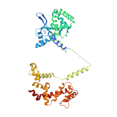
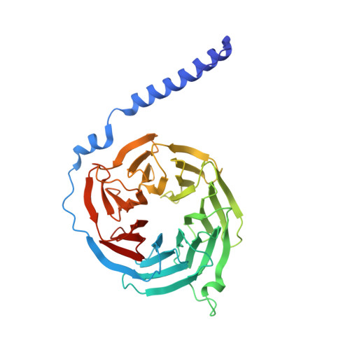
Structural organization of a major neuronal G protein regulator, the RGS7-G beta 5-R7BP complex.

Patil, D.N.Rangarajan, E.S.Novick, S.J.Pascal, B.D.Kojetin, D.J.Griffin, P.R.Izard, T.Martemyanov, K.A.

(2018) Elife 7

  • DOI: https://doi.org/10.7554/eLife.42150
  • Primary Citation of Related Structures:  
    6N9G

  • PubMed Abstract: 

    Signaling by the G-protein-coupled receptors (GPCRs) plays fundamental role in a vast number of essential physiological functions. Precise control of GPCR signaling requires action of regulators of G protein signaling (RGS) proteins that deactivate heterotrimeric G proteins. RGS proteins are elaborately regulated and comprise multiple domains and subunits, yet structural organization of these assemblies is poorly understood. Here, we report a crystal structure and dynamics analyses of the multisubunit complex of RGS7, a major regulator of neuronal signaling with key roles in controlling a number of drug target GPCRs and links to neuropsychiatric disease, metabolism, and cancer. The crystal structure in combination with molecular dynamics and mass spectrometry analyses reveals unique organizational features of the complex and long-range conformational changes imposed by its constituent subunits during allosteric modulation. Notably, several intermolecular interfaces in the complex work in synergy to provide coordinated modulation of this key GPCR regulator.


  • Organizational Affiliation

    Department of Neuroscience, The Scripps Research Institute, Jupiter, United States.


Macromolecules
Find similar proteins by:  (by identity cutoff)  |  3D Structure
Entity ID: 1
MoleculeChains Sequence LengthOrganismDetailsImage
Regulator of G-protein signaling 7
A, B
469Bos taurusMutation(s): 0 
Gene Names: RGS7
UniProt
Find proteins for O46470 (Bos taurus)
Explore O46470 
Go to UniProtKB:  O46470
Entity Groups  
Sequence Clusters30% Identity50% Identity70% Identity90% Identity95% Identity100% Identity
UniProt GroupO46470
Sequence Annotations
Expand
  • Reference Sequence
Find similar proteins by:  (by identity cutoff)  |  3D Structure
Entity ID: 2
MoleculeChains Sequence LengthOrganismDetailsImage
Guanine nucleotide-binding protein subunit beta-5
C, D
353Mus musculusMutation(s): 0 
Gene Names: Gnb5
UniProt & NIH Common Fund Data Resources
Find proteins for P62881 (Mus musculus)
Explore P62881 
Go to UniProtKB:  P62881
IMPC:  MGI:101848
Entity Groups  
Sequence Clusters30% Identity50% Identity70% Identity90% Identity95% Identity100% Identity
UniProt GroupP62881
Sequence Annotations
Expand
  • Reference Sequence
Experimental Data & Validation

Experimental Data

  • Method: X-RAY DIFFRACTION
  • Resolution: 2.13 Å
  • R-Value Free: 0.217 
  • R-Value Work: 0.174 
  • R-Value Observed: 0.176 
  • Space Group: P 1 21 1
Unit Cell:
Length ( Å )Angle ( ˚ )
a = 69.323α = 90
b = 162.76β = 98.36
c = 95.295γ = 90
Software Package:
Software NamePurpose
PHENIXrefinement
XDSdata reduction
autoPROCdata scaling
PHASERphasing

Structure Validation

View Full Validation Report

Currently 6N9G does not have a validation slider image.


Entry History & Funding Information

Deposition Data


Funding OrganizationLocationGrant Number
National Institutes of Health/National Institute on Drug Abuse (NIH/NIDA)United StatesDA036596

Revision History  (Full details and data files)

  • Version 1.0: 2019-01-09
    Type: Initial release
  • Version 1.1: 2019-12-11
    Changes: Author supporting evidence
  • Version 1.2: 2023-10-11
    Changes: Data collection, Database references, Refinement description