6MZP

Zebrafish betaglycan orphan domain structure from orthorhombic crystal form

  • Classification: PROTEIN BINDING
  • Organism(s): Danio rerio
  • Expression System: Homo sapiens
  • Mutation(s): No 

  • Deposited: 2018-11-05 Released: 2019-08-21 
  • Deposition Author(s): Hinck, A.P., Kim, S.
  • Funding Organization(s): National Institutes of Health/National Institute of General Medical Sciences (NIH/NIGMS), National Institutes of Health/National Cancer Institute (NIH/NCI)

Experimental Data Snapshot

  • Method: X-RAY DIFFRACTION
  • Resolution: 2.10 Å
  • R-Value Free: 0.258 
  • R-Value Work: 0.232 
  • R-Value Observed: 0.233 

wwPDB Validation   3D Report Full Report


This is version 1.2 of the entry. See complete history


Literature

Structural Adaptation in Its Orphan Domain Engenders Betaglycan with an Alternate Mode of Growth Factor Binding Relative to Endoglin.

Kim, S.K.Whitley, M.J.Krzysiak, T.C.Hinck, C.S.Taylor, A.B.Zwieb, C.Byeon, C.H.Zhou, X.Mendoza, V.Lopez-Casillas, F.Furey, W.Hinck, A.P.

(2019) Structure 27: 1427-1442.e4

  • DOI: https://doi.org/10.1016/j.str.2019.06.010
  • Primary Citation of Related Structures:  
    6MZN, 6MZP

  • PubMed Abstract: 

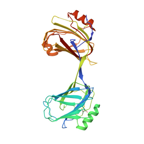
    Betaglycan (BG) and endoglin (ENG), homologous co-receptors of the TGF-β family, potentiate the signaling activity of TGF-β2 and inhibin A, and BMP-9 and BMP-10, respectively. BG exists as monomer and forms 1:1 growth factor (GF) complexes, while ENG exists as a dimer and forms 2:1 GF complexes. Herein, the structure of the BG orphan domain (BG O ) reveals an insertion that blocks the region that the endoglin orphan domain (ENG O ) uses to bind BMP-9, preventing it from binding in the same manner. Using binding studies with domain-deleted forms of TGF-β and BG O , as well as small-angle X-ray scattering data, BG O is shown to bind its cognate GF in an entirely different manner compared with ENG O . The alternative interfaces likely engender BG and ENG with the ability to selectively bind and target their cognate GFs in a unique temporal-spatial manner, without interfering with one another or other TGF-β family GFs.


  • Organizational Affiliation

    Department of Structural Biology, University of Pittsburgh School of Medicine, Biomedical Science Tower 3, Room 2051, 3501 Fifth Avenue, Pittsburgh, PA 15260, USA; Department of Biochemistry and Structural Biology, University of Texas Health Science Center at San Antonio, San Antonio, TX 78229-3900, USA.


Macromolecules
Find similar proteins by:  (by identity cutoff)  |  3D Structure
Entity ID: 1
MoleculeChains Sequence LengthOrganismDetailsImage
Transforming growth factor beta receptor III
A, B
338Danio rerioMutation(s): 0 
Gene Names: tgfbr3
UniProt
Find proteins for E7FDB6 (Danio rerio)
Explore E7FDB6 
Go to UniProtKB:  E7FDB6
Entity Groups  
Sequence Clusters30% Identity50% Identity70% Identity90% Identity95% Identity100% Identity
UniProt GroupE7FDB6
Sequence Annotations
Expand
  • Reference Sequence
Experimental Data & Validation

Experimental Data

  • Method: X-RAY DIFFRACTION
  • Resolution: 2.10 Å
  • R-Value Free: 0.258 
  • R-Value Work: 0.232 
  • R-Value Observed: 0.233 
  • Space Group: P 21 21 21
Unit Cell:
Length ( Å )Angle ( ˚ )
a = 63.52α = 90
b = 107.27β = 90
c = 113.24γ = 90
Software Package:
Software NamePurpose
BUSTERrefinement
MOSFLMdata reduction
SCALAdata scaling
SHARPphasing

Structure Validation

View Full Validation Report


Entry History & Funding Information

Deposition Data


Funding OrganizationLocationGrant Number
National Institutes of Health/National Institute of General Medical Sciences (NIH/NIGMS)United StatesGM58670
National Institutes of Health/National Cancer Institute (NIH/NCI)United StatesCA172886

Revision History  (Full details and data files)

  • Version 1.0: 2019-08-21
    Type: Initial release
  • Version 1.1: 2019-09-18
    Changes: Data collection, Database references
  • Version 1.2: 2019-12-04
    Changes: Author supporting evidence