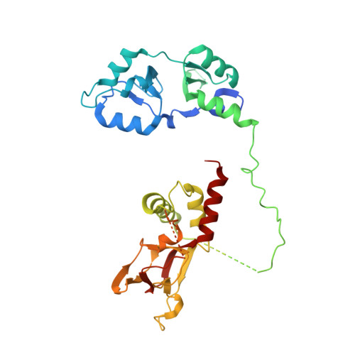
6MN6

Crystal structure of the cytosolic domain of human CNNM3


Experimental Data Snapshot

  • Method: X-RAY DIFFRACTION
  • Resolution: 3.36 Å
  • R-Value Free: 0.263 
  • R-Value Work: 0.217 

wwPDB Validation   3D Report Full Report

Currently 6MN6 does not have a validation slider image.


This is version 1.4 of the entry. See complete history


Literature

Mg2+-ATP Sensing in CNNM, a Putative Magnesium Transporter.

Chen, Y.S.Kozlov, G.Fakih, R.Yang, M.Zhang, Z.Kovrigin, E.L.Gehring, K.

(2020) Structure 28: 324-335.e4

  • DOI: https://doi.org/10.1016/j.str.2019.11.016
  • Primary Citation of Related Structures:  
    6MN6, 6N7E

  • PubMed Abstract: 

    The family of cystathionine-β-synthase (CBS)-pair domain divalent metal cation transport mediators (CNNMs) is composed of four integral membrane proteins associated with Mg 2+ transport. Structurally, CNNMs contain large cytosolic regions composed of a CBS-pair and a cyclic nucleotide-binding homology (CNBH) domain. How these regulate Mg 2+ transport activity is unknown. Here, we determined the crystal structures of cytosolic fragments in two conformations: Mg 2+ -ATP-analog bound and ligand free. The structures reveal open and closed conformations with functionally important contacts not observed in structures of the individual domains. We also identified a second Mg 2+ -binding region in the CBS-pair domain and a different dimerization interface for the CNBH domain. Analytical ultracentrifugation and isothermal titration calorimetry experiments revealed a tight correlation between Mg 2+ -ATP binding and protein dimerization. Mutations that blocked either function prevented cellular Mg 2+ efflux activity. The results suggest Mg 2+ efflux is regulated by conformational changes associated with Mg 2+ -ATP binding to CNNM CBS-pair domains.


  • Organizational Affiliation

    Department of Biochemistry & Centre for Structural Biology, McGill University, Montreal, QC H3G 0B1, Canada.


Macromolecules
Find similar proteins by:  (by identity cutoff)  |  3D Structure
Entity ID: 1
MoleculeChains Sequence LengthOrganismDetailsImage
Metal transporter CNNM3
A, B
365Homo sapiensMutation(s): 0 
Gene Names: CNNM3ACDP3
UniProt & NIH Common Fund Data Resources
Find proteins for Q8NE01 (Homo sapiens)
Explore Q8NE01 
Go to UniProtKB:  Q8NE01
PHAROS:  Q8NE01
GTEx:  ENSG00000168763 
Entity Groups  
Sequence Clusters30% Identity50% Identity70% Identity90% Identity95% Identity100% Identity
UniProt GroupQ8NE01
Sequence Annotations
Expand
  • Reference Sequence
Experimental Data & Validation

Experimental Data

  • Method: X-RAY DIFFRACTION
  • Resolution: 3.36 Å
  • R-Value Free: 0.263 
  • R-Value Work: 0.217 
  • Space Group: P 32 2 1
Unit Cell:
Length ( Å )Angle ( ˚ )
a = 128.063α = 90
b = 128.063β = 90
c = 111.316γ = 120
Software Package:
Software NamePurpose
PHENIXrefinement
HKL-2000data reduction
HKL-2000data scaling
PHENIXphasing

Structure Validation

View Full Validation Report

Currently 6MN6 does not have a validation slider image.


Entry History 

Deposition Data

Revision History  (Full details and data files)

  • Version 1.0: 2019-10-09
    Type: Initial release
  • Version 1.1: 2019-12-04
    Changes: Database references
  • Version 1.2: 2020-01-01
    Changes: Database references
  • Version 1.3: 2020-03-18
    Changes: Database references
  • Version 1.4: 2023-10-11
    Changes: Data collection, Database references, Refinement description