6MKT

Photoactive Yellow Protein with 3-chlorotyrosine substituted at position 42


Experimental Data Snapshot

  • Method: X-RAY DIFFRACTION
  • Resolution: 1.60 Å
  • R-Value Free: 0.189 
  • R-Value Work: 0.167 
  • R-Value Observed: 0.168 

wwPDB Validation   3D Report Full Report


Ligand Structure Quality Assessment 


This is version 1.4 of the entry. See complete history


Literature

Perturbation of Short Hydrogen Bonds in Photoactive Yellow Protein via Noncanonical Amino Acid Incorporation.

Thomson, B.Both, J.Wu, Y.Parrish, R.M.Martinez, T.J.Boxer, S.G.

(2019) J Phys Chem B 123: 4844-4849

  • DOI: https://doi.org/10.1021/acs.jpcb.9b01571
  • Primary Citation of Related Structures:  
    6MHI, 6MHN, 6MKT, 6MMD

  • PubMed Abstract: 

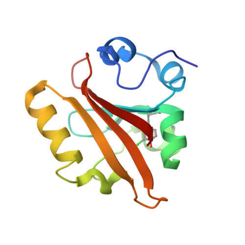
    Photoactive yellow protein (PYP) is a small photoreceptor protein that has two unusually short hydrogen bonds between the deprotonated p-coumaric acid chromophore and two amino acids, a tyrosine and a glutamic acid. This has led to considerable debate as to whether the glutamic acid-chromophore hydrogen bond is a low barrier hydrogen bond, with conflicting results in the literature. We have modified the p K a of the tyrosine by amber suppression and of the chromophore by chemical substitution. X-ray crystal structures of these modified proteins are nearly identical to the wild-type protein, so the heavy atom distance between proton donor and acceptor is maintained, even though these modifications change the relative proton affinity between donor and acceptor. Despite a considerable change in relative proton affinity, the NMR chemical shifts of the hydrogen-bonded protons are only moderately affected. QM/MM calculations were used to explore the protons' potential energy surface and connect the calculated proton position with empirically measured proton chemical shifts. The results are inconsistent with a low barrier hydrogen bond but in all cases are consistent with a localized proton, suggesting an ionic hydrogen bond rather than a low barrier hydrogen bond.


  • Organizational Affiliation

    Department of Chemistry , Stanford University , Stanford , California 94305 , United States.


Macromolecules
Find similar proteins by:  (by identity cutoff)  |  3D Structure
Entity ID: 1
MoleculeChains Sequence LengthOrganismDetailsImage
Photoactive yellow protein125Halorhodospira halophilaMutation(s): 0 
Gene Names: pyp
UniProt
Find proteins for P16113 (Halorhodospira halophila)
Explore P16113 
Go to UniProtKB:  P16113
Entity Groups  
Sequence Clusters30% Identity50% Identity70% Identity90% Identity95% Identity100% Identity
UniProt GroupP16113
Sequence Annotations
Expand
  • Reference Sequence
Small Molecules
Ligands 1 Unique
IDChains Name / Formula / InChI Key2D Diagram3D Interactions
HC4
Query on HC4

Download Ideal Coordinates CCD File 
B [auth A]4'-HYDROXYCINNAMIC ACID
C9 H8 O3
NGSWKAQJJWESNS-ZZXKWVIFSA-N
Modified Residues  1 Unique
IDChains TypeFormula2D DiagramParent
3CT
Query on 3CT
A
L-PEPTIDE LINKINGC9 H10 Cl N O3TYR
Experimental Data & Validation

Experimental Data

  • Method: X-RAY DIFFRACTION
  • Resolution: 1.60 Å
  • R-Value Free: 0.189 
  • R-Value Work: 0.167 
  • R-Value Observed: 0.168 
  • Space Group: P 63
Unit Cell:
Length ( Å )Angle ( ˚ )
a = 65.5α = 90
b = 65.5β = 90
c = 40.38γ = 120
Software Package:
Software NamePurpose
PHENIXrefinement
MOSFLMdata reduction
PROTEUM PLUSdata scaling
PHASERphasing

Structure Validation

View Full Validation Report


Ligand Structure Quality Assessment 


Entry History & Funding Information

Deposition Data


Funding OrganizationLocationGrant Number
National Institutes of Health/Office of the DirectorUnited StatesR35GM11804401
National Science Foundation (NSF, United States)United StatesCHE-17406456

Revision History  (Full details and data files)

  • Version 1.0: 2019-05-29
    Type: Initial release
  • Version 1.1: 2019-06-05
    Changes: Data collection, Database references
  • Version 1.2: 2019-06-26
    Changes: Data collection, Database references
  • Version 1.3: 2019-11-27
    Changes: Author supporting evidence
  • Version 1.4: 2023-10-11
    Changes: Data collection, Database references, Refinement description