6L5G

Crystal structure of yak lactoperoxidase with disordered heme moiety at 2.50 A resolution


Experimental Data Snapshot

  • Method: X-RAY DIFFRACTION
  • Resolution: 2.50 Å
  • R-Value Free: 0.239 
  • R-Value Work: 0.185 

wwPDB Validation   3D Report Full Report


Ligand Structure Quality Assessment 


This is version 2.3 of the entry. See complete history


Literature

Potassium-induced partial inhibition of lactoperoxidase: structure of the complex of lactoperoxidase with potassium ion at 2.20 angstrom resolution.

Singh, P.K.Pandey, S.Rani, C.Ahmad, N.Viswanathan, V.Sharma, P.Kaur, P.Sharma, S.Singh, T.P.

(2021) J Biol Inorg Chem 26: 149-159

  • DOI: https://doi.org/10.1007/s00775-020-01844-6
  • Primary Citation of Related Structures:  
    6L32, 6L5G, 7C73, 7C74, 7C75, 7D52, 7DMR

  • PubMed Abstract: 

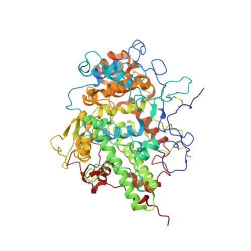
    Lactoperoxidase, a heme-containing glycoprotein, catalyzes the oxidation of thiocyanate by hydrogen peroxide into hypothiocyanite which acts as an antibacterial agent. The prosthetic heme moiety is attached to the protein through two ester linkages via Glu258 and Asp108. In lactoperoxidase, the substrate-binding site is formed on the distal heme side. To study the effect of physiologically important potassium ion on the structure and function of lactoperoxidase, the fresh protein samples were isolated from yak (Bos grunniens) colostrum and purified to homogeneity. The biochemical studies with potassium fluoride showed a significant reduction in the catalytic activity. Lactoperoxidase was crystallized using 200 mM ammonium nitrate and 20% PEG-3350 at pH 6.0. The crystals of LPO were soaked in the solution of potassium fluoride and used for the X-ray intensity data collection. Structure determination at 2.20 Å resolution revealed the presence of a potassium ion in the distal heme cavity. Structure determination further revealed that the propionic chain attached to pyrrole ring C of the heme moiety, was disordered into two components each having an occupancy of 0.5. One component occupied a position similar to the normally observed position of propionic chain while the second component was found in the distal heme cavity. The potassium ion in the distal heme cavity formed five coordinate bonds with two oxygen atoms of propionic moiety, N ε2 atom of His109 and two oxygen atoms of water molecules. The presence of potassium ion in the distal heme cavity hampered the catalytic activity of lactoperoxidase.


  • Organizational Affiliation

    Department of Biophysics, All India Institute of Medical Sciences, Ansari Nagar, New Delhi, 110 029, India.


Macromolecules
Find similar proteins by:  (by identity cutoff)  |  3D Structure
Entity ID: 1
MoleculeChains Sequence LengthOrganismDetailsImage
Lactoperoxidase595Bos mutusMutation(s): 0 
UniProt
Find proteins for P80025 (Bos taurus)
Explore P80025 
Go to UniProtKB:  P80025
Entity Groups  
Sequence Clusters30% Identity50% Identity70% Identity90% Identity95% Identity100% Identity
UniProt GroupP80025
Sequence Annotations
Expand
  • Reference Sequence
Oligosaccharides

Help

Entity ID: 2
MoleculeChains Length2D Diagram Glycosylation3D Interactions
2-acetamido-2-deoxy-beta-D-glucopyranose-(1-4)-2-acetamido-2-deoxy-beta-D-glucopyranose
B
2N-Glycosylation
Glycosylation Resources
GlyTouCan:  G42666HT
GlyCosmos:  G42666HT
GlyGen:  G42666HT
Small Molecules
Ligands 6 Unique
IDChains Name / Formula / InChI Key2D Diagram3D Interactions
HEM (Subject of Investigation/LOI)
Query on HEM

Download Ideal Coordinates CCD File 
G [auth A]PROTOPORPHYRIN IX CONTAINING FE
C34 H32 Fe N4 O4
KABFMIBPWCXCRK-RGGAHWMASA-L
NAG (Subject of Investigation/LOI)
Query on NAG

Download Ideal Coordinates CCD File 
C [auth A],
D [auth A],
E [auth A]
2-acetamido-2-deoxy-beta-D-glucopyranose
C8 H15 N O6
OVRNDRQMDRJTHS-FMDGEEDCSA-N
GOL
Query on GOL

Download Ideal Coordinates CCD File 
K [auth A],
L [auth A],
M [auth A],
N [auth A],
O [auth A]
GLYCEROL
C3 H8 O3
PEDCQBHIVMGVHV-UHFFFAOYSA-N
ZN
Query on ZN

Download Ideal Coordinates CCD File 
J [auth A]ZINC ION
Zn
PTFCDOFLOPIGGS-UHFFFAOYSA-N
CA
Query on CA

Download Ideal Coordinates CCD File 
F [auth A]CALCIUM ION
Ca
BHPQYMZQTOCNFJ-UHFFFAOYSA-N
K
Query on K

Download Ideal Coordinates CCD File 
H [auth A],
I [auth A]
POTASSIUM ION
K
NPYPAHLBTDXSSS-UHFFFAOYSA-N
Experimental Data & Validation

Experimental Data

  • Method: X-RAY DIFFRACTION
  • Resolution: 2.50 Å
  • R-Value Free: 0.239 
  • R-Value Work: 0.185 
  • Space Group: P 21 21 21
Unit Cell:
Length ( Å )Angle ( ˚ )
a = 79.82α = 90
b = 84.71β = 90
c = 99.04γ = 90
Software Package:
Software NamePurpose
REFMACrefinement
iMOSFLMdata reduction
Aimlessdata scaling
MOLREPphasing

Structure Validation

View Full Validation Report


Ligand Structure Quality Assessment 


Entry History 

Deposition Data

Revision History  (Full details and data files)

  • Version 1.0: 2019-11-06
    Type: Initial release
  • Version 2.0: 2020-07-29
    Type: Remediation
    Reason: Carbohydrate remediation
    Changes: Atomic model, Data collection, Derived calculations, Structure summary
  • Version 2.1: 2021-02-03
    Changes: Database references, Structure summary
  • Version 2.2: 2021-04-21
    Changes: Database references
  • Version 2.3: 2023-11-22
    Changes: Data collection, Database references, Derived calculations, Refinement description