Structural basis for the unusual substrate specificity of unique two-domain M1 metallopeptidase.
Agrawal, R., Goyal, V.D., Singh, R., Kumar, A., Jamdar, S.N., Kumar, A., Makde, R.D.(2020) Int J Biol Macromol 147: 304-313
- PubMed: 31923495
- DOI: https://doi.org/10.1016/j.ijbiomac.2019.12.239
- Primary Citation of Related Structures:
6IFF, 6KOY, 6KOZ, 6KP0, 6KP1 - PubMed Abstract:
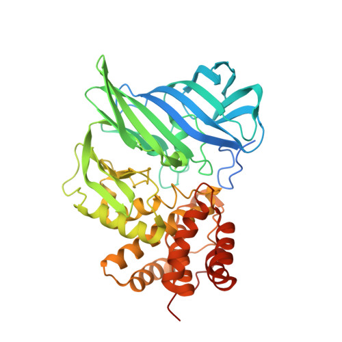
M1 metallopeptidases regulate many important biological processes such as angiogenesis, tumour growth, hormone regulation, and immune cell development. Knowledge of substrate specificity mechanism in this family is valuable. An M1 peptidase from Deinococcus radiodurans (M1dr) with preference for bulky hydrophobic residues at N-terminus of peptide substrates was recently reported. In contrast to Escherichia coli aminopeptidase N, a previously characterized M1 peptidase, M1dr exhibits reduced activity towards peptides with N-terminal Arg or Ala residue. In order to illuminate structural basis of substrate specificity, we report several crystal structures of M1dr with different amino acids bound to the active site. Structural analysis indicated that the enzyme makes subtle adjustments to multiple residues leading to significant volume change of the active site cavity to accommodate residues of varying sizes (Leu to Trp). This study further reveals that the low preference for Arg at N-terminus of peptide substrate arises from a non-productive conformation in which many of the Arg molecules bind where they block the proton donor essential for the peptidase reaction. Hence, this study illuminates the substrate-binding mechanism and also reveals the structural basis for the substrate specificity of M1dr enzyme.
Organizational Affiliation:
High Pressure and Synchrotron Radiation Physics Division, Bhabha Atomic Research Centre, Mumbai 400085, India; Discipline of Biosciences and Biomedical Engineering, Indian Institute of Technology Indore, Indore 453552, India.