


Crystal structure of the complex of phosphopantetheine adenylyl transferase from Acinetobacter baumannii with Dephospho Coenzyme at 3.2 A resolution.
Viswanathan, V., Gupta, A., Sharma, S., Singh, T.P.To be published.
Experimental Data Snapshot
Entity ID: 1 | |||||
|---|---|---|---|---|---|
| Molecule | Chains | Sequence Length | Organism | Details | Image |
| Phosphopantetheine adenylyltransferase | 163 | Acinetobacter baumannii | Mutation(s): 0 Gene Names: coaD EC: 2.7.7.3 | 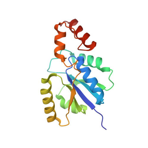 | |
UniProt | |||||
Find proteins for B0V8I3 (Acinetobacter baumannii (strain AYE)) Explore B0V8I3 Go to UniProtKB: B0V8I3 | |||||
Entity Groups | |||||
| Sequence Clusters | 30% Identity50% Identity70% Identity90% Identity95% Identity100% Identity | ||||
| UniProt Group | B0V8I3 | ||||
Sequence AnnotationsExpand | |||||
| |||||
| Ligands 2 Unique | |||||
|---|---|---|---|---|---|
| ID | Chains | Name / Formula / InChI Key | 2D Diagram | 3D Interactions | |
| COD (Subject of Investigation/LOI) Query on COD | C [auth A] | DEPHOSPHO COENZYME A C21 H35 N7 O13 P2 S KDTSHFARGAKYJN-IBOSZNHHSA-N |  | ||
| CL (Subject of Investigation/LOI) Query on CL | B [auth A] | CHLORIDE ION Cl VEXZGXHMUGYJMC-UHFFFAOYSA-M |  | ||
| Length ( Å ) | Angle ( ˚ ) |
|---|---|
| a = 215.539 | α = 90 |
| b = 215.539 | β = 90 |
| c = 215.539 | γ = 90 |
| Software Name | Purpose |
|---|---|
| REFMAC | refinement |
| XDS | data reduction |
| XSCALE | data scaling |
| MOLREP | phasing |
RCSB PDB (citation) is hosted by
RCSB PDB is a member of the

