6JUK

Crystal structure of Formate dehydrogenase mutant C256I/E261P/S381I from Pseudomonas sp. 101 in complex with non-natural cofactor Nicotinamide Cytosine Dinucleotide


Experimental Data Snapshot

  • Method: X-RAY DIFFRACTION
  • Resolution: 2.29 Å
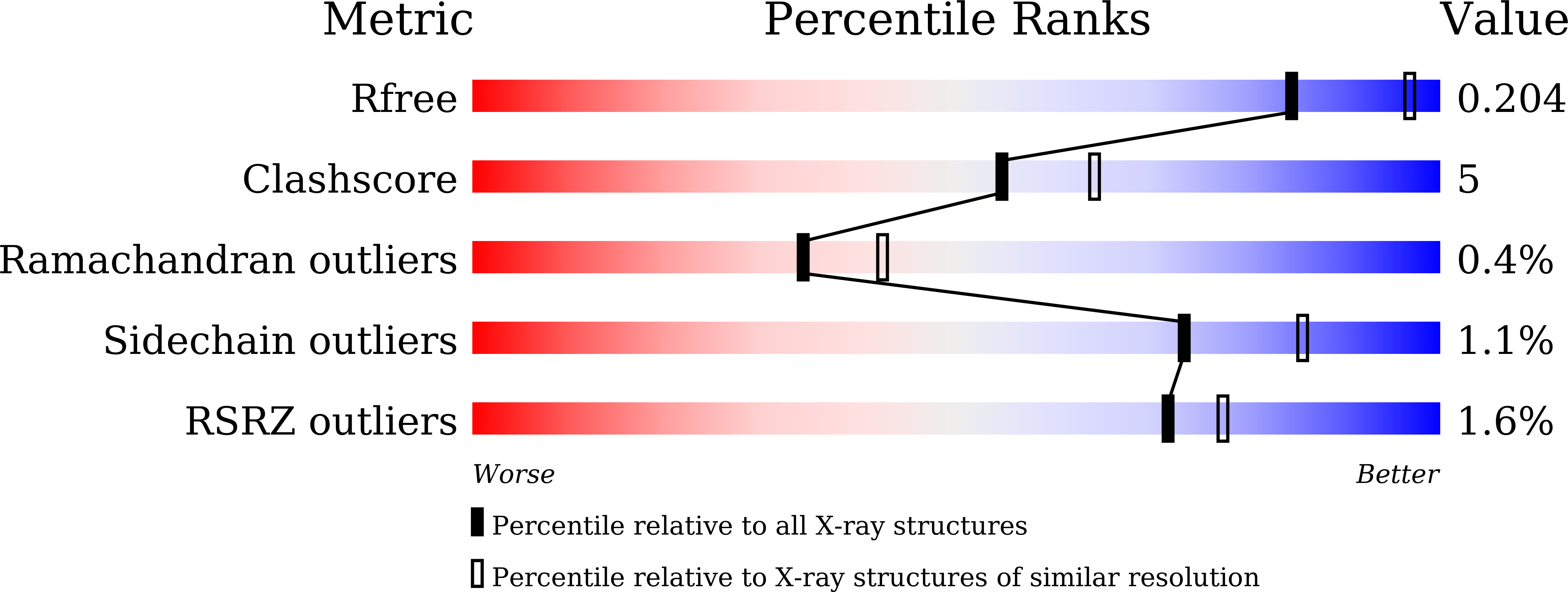
  • R-Value Free: 0.205 
  • R-Value Work: 0.159 
  • R-Value Observed: 0.161 

wwPDB Validation   3D Report Full Report


Ligand Structure Quality Assessment 


This is version 1.3 of the entry. See complete history


Literature

Structure-Guided Design of Formate Dehydrogenase for Regeneration of a Non-Natural Redox Cofactor.

Guo, X.Wang, X.Liu, Y.Li, Q.Wang, J.Liu, W.Zhao, Z.K.

(2020) Chemistry 26: 16611-16615

  • DOI: https://doi.org/10.1002/chem.202003102
  • Primary Citation of Related Structures:  
    6JUJ, 6JUK, 6JWG, 6JX1

  • PubMed Abstract: 

    Formate dehydrogenase (FDH) has been widely used for the regeneration of the reduced nicotinamide adenine dinucleotide (NADH). To utilize nicotinamide cytosine dinucleotide (NCD) as a non-natural redox cofactor, it remains challenging as NCDH, the reduced form of NCD, has to be efficiently regenerated. Here we demonstrate successful engineering of FDH for NCDH regeneration. Guided by the structural information of FDH from Pseudomonas sp. 101 (pseFDH) and the NAD-pseFDH complex, semi-rational strategies were applied to design mutant libraries and screen for NCD-linked activity. The most active mutant reached a cofactor preference switch from NAD to NCD by 3700-fold. Homology modeling analysis showed that these mutants had reduced cofactor binding pockets and dedicated hydrophobic interactions for NCD. Efficient regeneration of NCDH was implemented by powering an NCD-dependent D-lactate dehydrogenase for stoichiometric and stereospecific reduction of pyruvate to D-lactate at the expense of formate.


  • Organizational Affiliation

    Laboratory of Biotechnology, Dalian Institute of Chemical Physics, Chinese Academy of Sciences, Dalian, 116023, P. R. China.


Macromolecules
Find similar proteins by:  (by identity cutoff)  |  3D Structure
Entity ID: 1
MoleculeChains Sequence LengthOrganismDetailsImage
Formate dehydrogenase
A, B
401Pseudomonas sp. 101Mutation(s): 3 
EC: 1.17.1.9
UniProt
Find proteins for P33160 (Pseudomonas sp. (strain 101))
Explore P33160 
Go to UniProtKB:  P33160
Entity Groups  
Sequence Clusters30% Identity50% Identity70% Identity90% Identity95% Identity100% Identity
UniProt GroupP33160
Sequence Annotations
Expand
  • Reference Sequence
Small Molecules
Ligands 2 Unique
IDChains Name / Formula / InChI Key2D Diagram3D Interactions
A7R
Query on A7R

Download Ideal Coordinates CCD File 
G [auth A],
H [auth B]
[[(2S,3S,4R,5S)-5-(3-aminocarbonylpyridin-1-ium-1-yl)-3,4-bis(oxidanyl)oxolan-2-yl]methoxy-oxidanyl-phosphoryl] [(2S,3S,4R,5S)-5-(4-azanyl-2-oxidanylidene-pyrimidin-1-yl)-3,4-bis(oxidanyl)oxolan-2-yl]methyl hydrogen phosphate
C20 H28 N5 O15 P2
CRWNYXSXULHVKY-JKHXZYGESA-O
GOL
Query on GOL

Download Ideal Coordinates CCD File 
C [auth A]
D [auth A]
E [auth A]
F [auth A]
I [auth B]
C [auth A],
D [auth A],
E [auth A],
F [auth A],
I [auth B],
J [auth B]
GLYCEROL
C3 H8 O3
PEDCQBHIVMGVHV-UHFFFAOYSA-N
Experimental Data & Validation

Experimental Data

  • Method: X-RAY DIFFRACTION
  • Resolution: 2.29 Å
  • R-Value Free: 0.205 
  • R-Value Work: 0.159 
  • R-Value Observed: 0.161 
  • Space Group: P 1 21 1
Unit Cell:
Length ( Å )Angle ( ˚ )
a = 78.038α = 90
b = 54.181β = 99.72
c = 102.828γ = 90
Software Package:
Software NamePurpose
PHENIXrefinement
HKL-2000data reduction
HKL-2000data scaling
PHENIXphasing

Structure Validation

View Full Validation Report


Ligand Structure Quality Assessment 


Entry History 

Deposition Data

Revision History  (Full details and data files)

  • Version 1.0: 2020-05-13
    Type: Initial release
  • Version 1.1: 2020-12-02
    Changes: Database references
  • Version 1.2: 2020-12-30
    Changes: Database references
  • Version 1.3: 2023-11-22
    Changes: Data collection, Database references, Refinement description