6JKU

Crystal structure of N-acetylglucosamine-6-phosphate deacetylase from Pasteurella Multocida


Experimental Data Snapshot

  • Method: X-RAY DIFFRACTION
  • Resolution: 1.95 Å
  • R-Value Free: 0.200 
  • R-Value Work: 0.163 
  • R-Value Observed: 0.165 

wwPDB Validation   3D Report Full Report


This is version 1.3 of the entry. See complete history


Literature

Quaternary variations in the structural assembly of N-acetylglucosamine-6-phosphate deacetylase from Pasteurella multocida.

Manjunath, L.Coombes, D.Davies, J.Dhurandhar, M.Tiwari, V.R.Dobson, R.C.J.Sowdhamini, R.Ramaswamy, S.Bose, S.

(2020) Proteins 

  • DOI: https://doi.org/10.1002/prot.25996
  • Primary Citation of Related Structures:  
    6JKU

  • PubMed Abstract: 

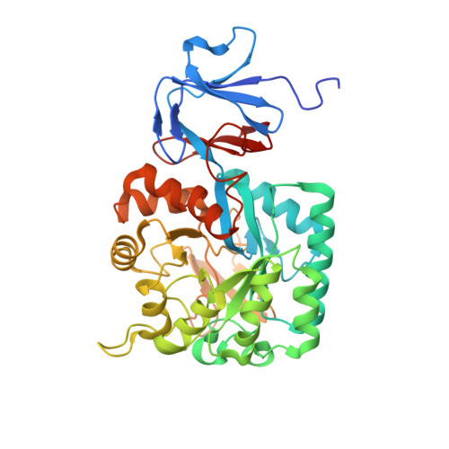
    N-acetylglucosamine 6-phosphate deacetylase (NagA) catalyzes the conversion of N-acetylglucosamine-6-phosphate to glucosamine-6-phosphate in amino sugar catabolism. This conversion is an essential step in the catabolism of sialic acid in several pathogenic bacteria, including Pasteurella multocida, and thus NagA is identified as a potential drug target. Here, we report the unique structural features of NagA from P. multocida (PmNagA) resolved to 1.95 Å. PmNagA displays an altered quaternary architecture with unique interface interactions compared to its close homolog, the Escherichia coli NagA (EcNagA). We confirmed that the altered quaternary structure is not a crystallographic artifact using single particle electron cryo-microscopy. Analysis of the determined crystal structure reveals a set of hot-spot residues involved in novel interactions at the dimer-dimer interface. PmNagA binds to one Zn 2+ ion in the active site and demonstrates kinetic parameters comparable to other bacterial homologs. Kinetic studies reveal that at high substrate concentrations (~10-fold the K M ), the tetrameric PmNagA displays hysteresis similar to its distant neighbor, the dimeric Staphylococcus aureus NagA (SaNagA). Our findings provide key information on structural and functional properties of NagA in P. multocida that could be utilized to design novel antibacterials.


  • Organizational Affiliation

    Institute for Stem Cell Science and Regenerative Medicine, NCBS, GKVK Campus, Bangalore, Karnataka, India.


Macromolecules
Find similar proteins by:  (by identity cutoff)  |  3D Structure
Entity ID: 1
MoleculeChains Sequence LengthOrganismDetailsImage
N-acetylglucosamine-6-phosphate deacetylase
A, B, C, D
395Pasteurella multocidaMutation(s): 0 
Gene Names: BGK37_09560
UniProt
Find proteins for Q9CMF5 (Pasteurella multocida (strain Pm70))
Explore Q9CMF5 
Go to UniProtKB:  Q9CMF5
Entity Groups  
Sequence Clusters30% Identity50% Identity70% Identity90% Identity95% Identity100% Identity
UniProt GroupQ9CMF5
Sequence Annotations
Expand
  • Reference Sequence
Small Molecules
Ligands 5 Unique
IDChains Name / Formula / InChI Key2D Diagram3D Interactions
PO4
Query on PO4

Download Ideal Coordinates CCD File 
F [auth A]
G [auth A]
GA [auth C]
H [auth A]
HA [auth C]
F [auth A],
G [auth A],
GA [auth C],
H [auth A],
HA [auth C],
I [auth A],
IA [auth C],
J [auth A],
PA [auth D],
QA [auth D],
RA [auth D],
SA [auth D],
U [auth B],
V [auth B],
W [auth B]
PHOSPHATE ION
O4 P
NBIIXXVUZAFLBC-UHFFFAOYSA-K
GOL
Query on GOL

Download Ideal Coordinates CCD File 
DA [auth B],
R [auth A]
GLYCEROL
C3 H8 O3
PEDCQBHIVMGVHV-UHFFFAOYSA-N
ZN
Query on ZN

Download Ideal Coordinates CCD File 
E [auth A],
FA [auth C],
OA [auth D],
T [auth B]
ZINC ION
Zn
PTFCDOFLOPIGGS-UHFFFAOYSA-N
EDO
Query on EDO

Download Ideal Coordinates CCD File 
AA [auth B]
BA [auth B]
CA [auth B]
JA [auth C]
K [auth A]
AA [auth B],
BA [auth B],
CA [auth B],
JA [auth C],
K [auth A],
KA [auth C],
L [auth A],
LA [auth C],
M [auth A],
MA [auth C],
N [auth A],
O [auth A],
P [auth A],
Q [auth A],
TA [auth D],
UA [auth D],
VA [auth D],
WA [auth D],
X [auth B],
XA [auth D],
Y [auth B],
Z [auth B]
1,2-ETHANEDIOL
C2 H6 O2
LYCAIKOWRPUZTN-UHFFFAOYSA-N
NA
Query on NA

Download Ideal Coordinates CCD File 
EA [auth B],
NA [auth C],
S [auth A],
YA [auth D]
SODIUM ION
Na
FKNQFGJONOIPTF-UHFFFAOYSA-N
Experimental Data & Validation

Experimental Data

  • Method: X-RAY DIFFRACTION
  • Resolution: 1.95 Å
  • R-Value Free: 0.200 
  • R-Value Work: 0.163 
  • R-Value Observed: 0.165 
  • Space Group: P 21 21 21
Unit Cell:
Length ( Å )Angle ( ˚ )
a = 89.162α = 90
b = 139.549β = 90
c = 166.556γ = 90
Software Package:
Software NamePurpose
PHENIXrefinement
PDB_EXTRACTdata extraction
XDSdata reduction
Aimlessdata scaling
PHASERphasing

Structure Validation

View Full Validation Report


Entry History & Funding Information

Deposition Data


Funding OrganizationLocationGrant Number
Department of Biotechnology (India)IndiaBT/INF/22/SP22660/2017
Department of Biotechnology (India)IndiaBT/PR12422/MED /31/287/214
Department of Biotechnology (India)IndiaBT/PR5081/INF/156/2012
Department of Biotechnology (India)IndiaBT/IN/SWEDEN/06/SR/2017-2018

Revision History  (Full details and data files)

  • Version 1.0: 2020-03-04
    Type: Initial release
  • Version 1.1: 2020-04-08
    Changes: Derived calculations
  • Version 1.2: 2021-03-17
    Changes: Database references, Derived calculations
  • Version 1.3: 2023-11-22
    Changes: Data collection, Database references, Refinement description