


Actinonin bound crystal structure of class I type b peptide deformylase from Acinetobacter baumannii
Lee, I.H., Ho, T.H., Kang, L.W.To be published.
Experimental Data Snapshot
Entity ID: 1 | |||||
|---|---|---|---|---|---|
| Molecule | Chains | Sequence Length | Organism | Details | Image |
| Peptide deformylase | 159 | Acinetobacter baumannii | Mutation(s): 0 Gene Names: def, C3415_07350, IX87_00730 EC: 3.5.1.88 | 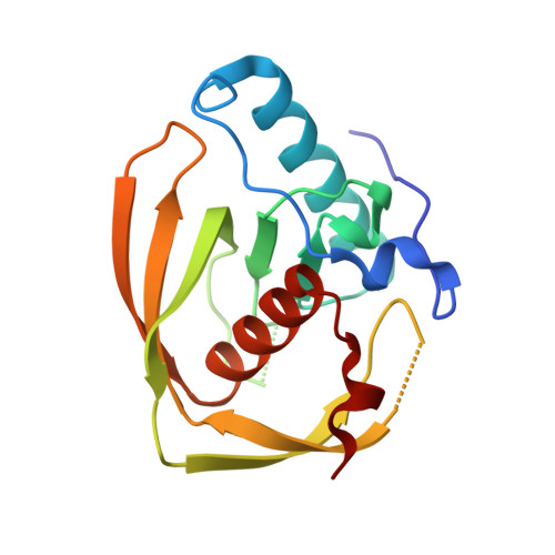 | |
Entity Groups | |||||
| Sequence Clusters | 30% Identity50% Identity70% Identity90% Identity95% Identity100% Identity | ||||
Sequence AnnotationsExpand | |||||
| |||||
| Ligands 2 Unique | |||||
|---|---|---|---|---|---|
| ID | Chains | Name / Formula / InChI Key | 2D Diagram | 3D Interactions | |
| BB2 (Subject of Investigation/LOI) Query on BB2 | D [auth A], F [auth B] | ACTINONIN C19 H35 N3 O5 XJLATMLVMSFZBN-VYDXJSESSA-N |  | ||
| ZN Query on ZN | C [auth A], E [auth B] | ZINC ION Zn PTFCDOFLOPIGGS-UHFFFAOYSA-N |  | ||
| Length ( Å ) | Angle ( ˚ ) |
|---|---|
| a = 39.728 | α = 90 |
| b = 70.786 | β = 90 |
| c = 110.859 | γ = 90 |
| Software Name | Purpose |
|---|---|
| HKL-2000 | data scaling |
| REFMAC | refinement |
| HKL-2000 | data collection |
| HKL-2000 | data reduction |
| MOLREP | phasing |
RCSB PDB (citation) is hosted by
RCSB PDB is a member of the

