3,6-Anhydro-L-Galactose Dehydrogenase VvAHGD is a Member of a New Aldehyde Dehydrogenase Family and Catalyzes by a Novel Mechanism with Conformational Switch of Two Catalytic Residues Cysteine 282 and Glutamate 248.
Wang, Y., Li, P.Y., Zhang, Y., Cao, H.Y., Wang, Y.J., Li, C.Y., Wang, P., Su, H.N., Chen, Y., Chen, X.L., Zhang, Y.Z.(2020) J Mol Biol 432: 2186-2203
- PubMed: 32087198
- DOI: https://doi.org/10.1016/j.jmb.2020.02.008
- Primary Citation of Related Structures:
6J75, 6J76 - PubMed Abstract:
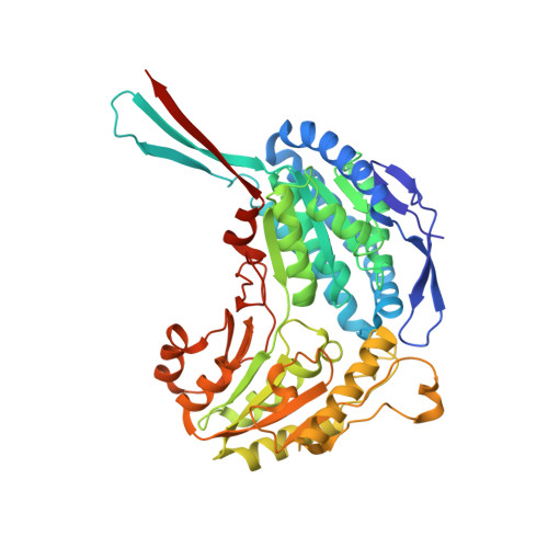
3,6-anhydro-α-L-galactose (L-AHG) is one of the main monosaccharide constituents of red macroalgae. In the recently discovered bacterial L-AHG catabolic pathway, L-AHG is first oxidized by a NAD(P) + -dependent dehydrogenase (AHGD), which is a key step of this pathway. However, the catalytic mechanism(s) of AHGDs is still unclear. Here, we identified and characterized an AHGD from marine bacterium Vibrio variabilis JCM 19239 (VvAHGD). The NADP + -dependent VvAHGD could efficiently oxidize L-AHG. Phylogenetic analysis suggested that VvAHGD and its homologs represent a new aldehyde dehydrogenase (ALDH) family with different substrate preferences from reported ALDH families, named the L-AHGDH family. To explain the catalytic mechanism of VvAHGD, we solved the structures of VvAHGD in the apo form and complex with NADP + and modeled its structure with L-AHG. Based on structural, mutational, and biochemical analyses, the cofactor channel and the substrate channel of VvAHGD are identified, and the key residues involved in the binding of NADP + and L-AHG and the catalysis are revealed. VvAHGD performs catalysis by controlling the consecutive connection and interruption of the cofactor channel and the substrate channel via the conformational changes of its two catalytic residues Cys282 and Glu248. Comparative analyses of structures and enzyme kinetics revealed that differences in the substrate channels (in shape, size, electrostatic surface, and residue composition) lead to the different substrate preferences of VvAHGD from other ALDHs. This study on VvAHGD sheds light on the diversified catalytic mechanisms and evolution of NAD(P) + -dependent ALDHs.
Organizational Affiliation:
State Key Laboratory of Microbial Technology, Marine Biotechnology Research Center, Shandong University, Qingdao, 266237, China.