6J07

Crystal structure of human TERB2 and TERB1


Experimental Data Snapshot

  • Method: X-RAY DIFFRACTION
  • Resolution: 3.30 Å
  • R-Value Free: 0.337 
  • R-Value Work: 0.288 
  • R-Value Observed: 0.291 

wwPDB Validation   3D Report Full Report


This is version 1.0 of the entry. See complete history


Literature

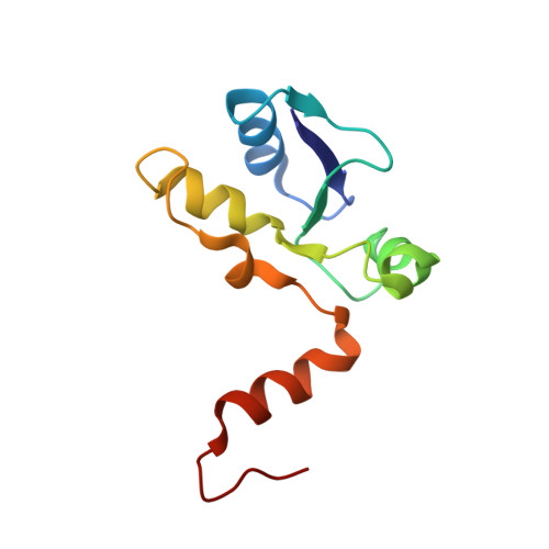
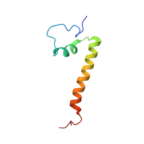
The meiotic TERB1-TERB2-MAJIN complex tethers telomeres to the nuclear envelope.

Wang, Y.Chen, Y.Chen, J.Wang, L.Nie, L.Long, J.Chang, H.Wu, J.Huang, C.Lei, M.

(2019) Nat Commun 10: 564-564

  • DOI: https://doi.org/10.1038/s41467-019-08437-1
  • Primary Citation of Related Structures:  
    6J07, 6J08

  • PubMed Abstract: 

    During meiotic prophase I, telomeres attach to and move on the nuclear envelope (NE), regulating chromosome movement to promote homologous pairing. Meiosis-specific proteins TERB1, TERB2 and MAJIN play a key role in this process. Here, we report the crystal structures of human TERB1-TERB2 and TERB2-MAJIN subcomplexes. Specific disruption of the TERB1-TERB2 or the TERB2-MAJIN interaction in the mouse Terb2 gene abolishes the telomere attachment to the NE and causes aberrant homologous pairing and disordered synapsis. In addition, depletion of SUN1 also partially disrupts the telomere-NE connection. We propose that the telomere-TRF1-TERB1-TERB2-MAJIN-NE interaction network and the telomere-LINC complex connection are likely two separate but cooperative pathways to stably recruit telomeres to the NE in meiosis prophase I. Our work provides a molecular model of the connection between telomeres and the NE and reveals the correlation between aberrant synapsis and the defective telomere attachment to the NE.


  • Organizational Affiliation

    National Center for Protein Science Shanghai, State Key Laboratory of Molecular Biology, CAS Center for Excellence on Molecular Cell Science, Institute of Biochemistry and Cell Biology, Chinese Academy of Sciences, University of Chinese Academy of Sciences, 200031, Shanghai, China.


Macromolecules
Find similar proteins by:  (by identity cutoff)  |  3D Structure
Entity ID: 1
MoleculeChains Sequence LengthOrganismDetailsImage
Telomere repeats-binding bouquet formation protein 2107Homo sapiensMutation(s): 0 
Gene Names: TERB2C15orf43
UniProt & NIH Common Fund Data Resources
Find proteins for Q8NHR7 (Homo sapiens)
Explore Q8NHR7 
Go to UniProtKB:  Q8NHR7
PHAROS:  Q8NHR7
GTEx:  ENSG00000167014 
Entity Groups  
Sequence Clusters30% Identity50% Identity70% Identity90% Identity95% Identity100% Identity
UniProt GroupQ8NHR7
Sequence Annotations
Expand
  • Reference Sequence
Find similar proteins by:  (by identity cutoff)  |  3D Structure
Entity ID: 2
MoleculeChains Sequence LengthOrganismDetailsImage
Telomere repeats-binding bouquet formation protein 160Homo sapiensMutation(s): 0 
Gene Names: TERB1CCDC79
UniProt & NIH Common Fund Data Resources
Find proteins for Q8NA31 (Homo sapiens)
Explore Q8NA31 
Go to UniProtKB:  Q8NA31
PHAROS:  Q8NA31
GTEx:  ENSG00000249961 
Entity Groups  
Sequence Clusters30% Identity50% Identity70% Identity90% Identity95% Identity100% Identity
UniProt GroupQ8NA31
Sequence Annotations
Expand
  • Reference Sequence
Small Molecules
Ligands 1 Unique
IDChains Name / Formula / InChI Key2D Diagram3D Interactions
ZN
Query on ZN

Download Ideal Coordinates CCD File 
C [auth B]ZINC ION
Zn
PTFCDOFLOPIGGS-UHFFFAOYSA-N
Experimental Data & Validation

Experimental Data

  • Method: X-RAY DIFFRACTION
  • Resolution: 3.30 Å
  • R-Value Free: 0.337 
  • R-Value Work: 0.288 
  • R-Value Observed: 0.291 
  • Space Group: P 64 2 2
Unit Cell:
Length ( Å )Angle ( ˚ )
a = 126.9α = 90
b = 126.9β = 90
c = 90.429γ = 120
Software Package:
Software NamePurpose
SCALEPACKdata scaling
PHENIXrefinement
PDB_EXTRACTdata extraction
HKL-3000data collection
PHENIXphasing

Structure Validation

View Full Validation Report


Entry History 

Deposition Data

Revision History  (Full details and data files)

  • Version 1.0: 2019-02-27
    Type: Initial release