6IU7

Crystal structure of importin-alpha1 bound to the 53BP1 nuclear localization signal (wild-type)


Experimental Data Snapshot

  • Method: X-RAY DIFFRACTION
  • Resolution: 1.90 Å
  • R-Value Free: 0.185 
  • R-Value Work: 0.153 
  • R-Value Observed: 0.155 

wwPDB Validation   3D Report Full Report


This is version 1.2 of the entry. See complete history


Literature

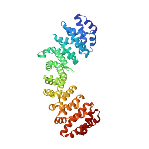
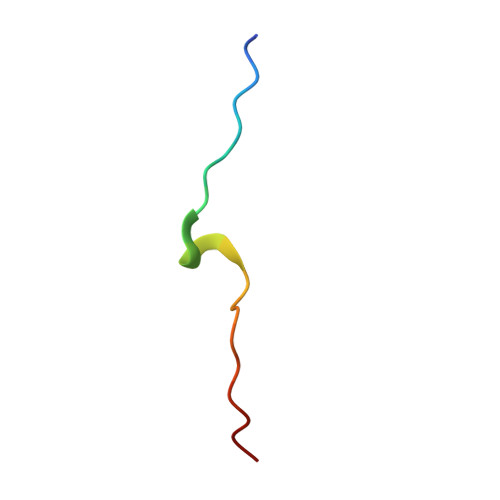
Structural and biochemical characterization of the recognition of the 53BP1 nuclear localization signal by importin-alpha.

Matsuura, Y.

(2019) Biochem Biophys Res Commun 510: 236-241

  • DOI: https://doi.org/10.1016/j.bbrc.2019.01.075
  • Primary Citation of Related Structures:  
    6IU7, 6IUA

  • PubMed Abstract: 

    53BP1 (TP53-binding protein 1) plays a key role in DNA double-strand break repair by promoting non-homologous end joining (NHEJ) especially during G1 phase of the cell cycle. Nuclear import of 53BP1 is required for proper localization of 53BP1 and maintenance of genome integrity. 53BP1 has a classical bipartite nuclear localization signal (NLS) of sequence 1666-GKRKLITSEEERSPAKRGRKS-1686. Ser1678 within the 53BP1 NLS can be phosphorylated by CDK1/cyclin B, and a phosphomimetic substitution of Ser1678 with aspartate has been shown to negatively regulate nuclear import of 53BP1. Here, the X-ray crystal structures of the nuclear import adaptor importin-α1 bound to the wild-type 53BP1 NLS and the S1678D mutant of 53BP1 NLS are reported at resolutions of 1.9 and 1.7 Å, respectively. In the wild-type structure, not only the two basic clusters of the 53BP1 NLS but also the linker region between the basic clusters made extensive interactions with importin-α1. In the mutant structure, the linker region between the basic clusters in the 53BP1 NLS made fewer interactions with importin-α1 than those observed in the wild-type structure. However, biochemical binding assays using purified proteins showed that the 53BP1 mutation S1678D reduces the binding affinity to importin-α1 only to a modest extent. Implications of these findings for regulatory mechanism of 53BP1 nuclear import are discussed.


  • Organizational Affiliation

    Division of Biological Science, Graduate School of Science, Nagoya University, Japan; Structural Biology Research Center, Graduate School of Science, Nagoya University, Japan. Electronic address: matsuura.yoshiyuki@d.mbox.nagoya-u.ac.jp.


Macromolecules
Find similar proteins by:  (by identity cutoff)  |  3D Structure
Entity ID: 1
MoleculeChains Sequence LengthOrganismDetailsImage
Importin subunit alpha-1427Mus musculusMutation(s): 0 
Gene Names: Kpna2Rch1
UniProt & NIH Common Fund Data Resources
Find proteins for P52293 (Mus musculus)
Explore P52293 
Go to UniProtKB:  P52293
IMPC:  MGI:103561
Entity Groups  
Sequence Clusters30% Identity50% Identity70% Identity90% Identity95% Identity100% Identity
UniProt GroupP52293
Sequence Annotations
Expand
  • Reference Sequence

Find similar proteins by:  Sequence   |   3D Structure  

Entity ID: 2
MoleculeChains Sequence LengthOrganismDetailsImage
Peptide from TP53-binding protein 122Homo sapiensMutation(s): 0 
Gene Names: TP53BP1
UniProt & NIH Common Fund Data Resources
Find proteins for Q12888 (Homo sapiens)
Explore Q12888 
Go to UniProtKB:  Q12888
PHAROS:  Q12888
GTEx:  ENSG00000067369 
Entity Groups  
Sequence Clusters30% Identity50% Identity70% Identity90% Identity95% Identity100% Identity
UniProt GroupQ12888
Sequence Annotations
Expand
  • Reference Sequence
Experimental Data & Validation

Experimental Data

  • Method: X-RAY DIFFRACTION
  • Resolution: 1.90 Å
  • R-Value Free: 0.185 
  • R-Value Work: 0.153 
  • R-Value Observed: 0.155 
  • Space Group: P 21 21 21
Unit Cell:
Length ( Å )Angle ( ˚ )
a = 79.46α = 90
b = 90.15β = 90
c = 96.72γ = 90
Software Package:
Software NamePurpose
Aimlessdata scaling
PHENIXrefinement
PDB_EXTRACTdata extraction
MOSFLMdata reduction
MOLREPphasing

Structure Validation

View Full Validation Report


Entry History 

Deposition Data

  • Released Date: 2019-01-30 
  • Deposition Author(s): Matsuura, Y.

Revision History  (Full details and data files)

  • Version 1.0: 2019-01-30
    Type: Initial release
  • Version 1.1: 2019-02-20
    Changes: Data collection, Database references
  • Version 1.2: 2023-11-22
    Changes: Data collection, Database references, Refinement description