6IQL

Crystal structure of dopamine receptor D4 bound to the subtype-selective ligand, L745870


Experimental Data Snapshot

  • Method: X-RAY DIFFRACTION
  • Resolution: 3.50 Å
  • R-Value Free: 0.336 
  • R-Value Work: 0.306 
  • R-Value Observed: 0.307 

wwPDB Validation   3D Report Full Report


Ligand Structure Quality Assessment 


This is version 1.1 of the entry. See complete history


Literature

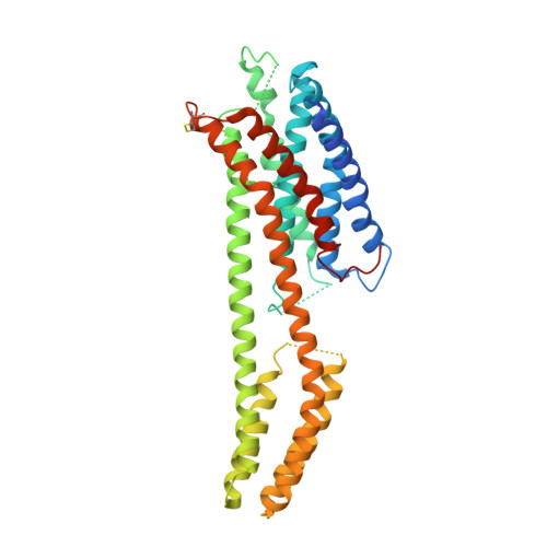
Crystal structure of dopamine receptor D4 bound to the subtype selective ligand, L745870.

Zhou, Y.Cao, C.He, L.Wang, X.Zhang, X.C.

(2019) Elife 8

  • DOI: https://doi.org/10.7554/eLife.48822
  • Primary Citation of Related Structures:  
    6IQL

  • PubMed Abstract: 

    Multiple subtypes of dopamine receptors within the GPCR superfamily regulate neurological processes through various downstream signaling pathways. A crucial question about the dopamine receptor family is what structural features determine the subtype-selectivity of potential drugs. Here, we report the 3.5-angstrom crystal structure of mouse dopamine receptor D4 (DRD4) complexed with a subtype-selective antagonist, L745870. Our structure reveals a secondary binding pocket extended from the orthosteric ligand-binding pocket to a DRD4-specific crevice located between transmembrane helices 2 and 3. Additional mutagenesis studies suggest that the antagonist L745870 prevents DRD4 activation by blocking the relative movement between transmembrane helices 2 and 3. These results expand our knowledge of the molecular basis for the physiological functions of DRD4 and assist new drug design.


  • Organizational Affiliation

    National Laboratory of Biomacromolecules, CAS Center for Excellence in Biomacromolecules, Institute of Biophysics, Chinese Academy of Sciences, Beijing, China.


Macromolecules
Find similar proteins by:  (by identity cutoff)  |  3D Structure
Entity ID: 1
MoleculeChains Sequence LengthOrganismDetailsImage
D(4) dopamine receptor,Soluble cytochrome b562,D(4) dopamine receptor
A, B
396Mus musculusEscherichia coliMutation(s): 6 
Gene Names: Drd4cybC
Membrane Entity: Yes 
UniProt
Find proteins for P0ABE7 (Escherichia coli)
Explore P0ABE7 
Go to UniProtKB:  P0ABE7
Find proteins for P51436 (Mus musculus)
Explore P51436 
Go to UniProtKB:  P51436
Entity Groups  
Sequence Clusters30% Identity50% Identity70% Identity90% Identity95% Identity100% Identity
UniProt GroupsP0ABE7P51436
Sequence Annotations
Expand
  • Reference Sequence
Small Molecules
Ligands 1 Unique
IDChains Name / Formula / InChI Key2D Diagram3D Interactions
L74 (Subject of Investigation/LOI)
Query on L74

Download Ideal Coordinates CCD File 
C [auth A],
D [auth B]
3-{[4-(4-chlorophenyl)piperazin-1-yl]methyl}-1H-pyrrolo[2,3-b]pyridine
C18 H19 Cl N4
OGJGQVFWEPNYSB-UHFFFAOYSA-N
Experimental Data & Validation

Experimental Data

  • Method: X-RAY DIFFRACTION
  • Resolution: 3.50 Å
  • R-Value Free: 0.336 
  • R-Value Work: 0.306 
  • R-Value Observed: 0.307 
  • Space Group: P 21 2 21
Unit Cell:
Length ( Å )Angle ( ˚ )
a = 46.7α = 90
b = 142β = 90
c = 146γ = 90
Software Package:
Software NamePurpose
PHENIXrefinement
XDSdata reduction
XSCALEdata scaling
PDB_EXTRACTdata extraction
PHASERphasing

Structure Validation

View Full Validation Report


Ligand Structure Quality Assessment 


Entry History & Funding Information

Deposition Data


Funding OrganizationLocationGrant Number
Ministry of Science and Technology (China)China2015CB910104
National Natural Science Foundation of ChinaChina31470745
Chinese Academy of SciencesChinaXDB08020301

Revision History  (Full details and data files)

  • Version 1.0: 2019-12-04
    Type: Initial release
  • Version 1.1: 2023-11-22
    Changes: Data collection, Database references, Refinement description