6IEJ

The C2 domain of cytosolic phospholipase A2 alpha bound to phosphatidylcholine


Experimental Data Snapshot

  • Method: X-RAY DIFFRACTION
  • Resolution: 2.21 Å
  • R-Value Free: 0.249 
  • R-Value Work: 0.224 
  • R-Value Observed: 0.226 

wwPDB Validation   3D Report Full Report


Ligand Structure Quality Assessment 


This is version 1.2 of the entry. See complete history


Literature

Structural basis of phosphatidylcholine recognition by the C2-domain of cytosolic phospholipase A2alpha.

Hirano, Y.Gao, Y.G.Stephenson, D.J.Vu, N.T.Malinina, L.Simanshu, D.K.Chalfant, C.E.Patel, D.J.Brown, R.E.

(2019) Elife 8

  • DOI: https://doi.org/10.7554/eLife.44760
  • Primary Citation of Related Structures:  
    6IEJ

  • PubMed Abstract: 

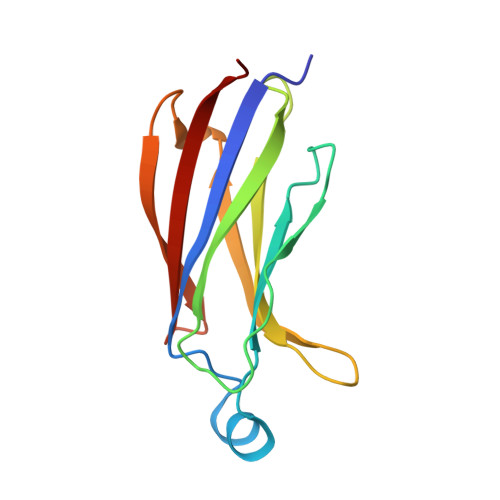
    Ca 2+ -stimulated translocation of cytosolic phospholipase A 2 α (cPLA 2 α) to the Golgi induces arachidonic acid production, the rate-limiting step in pro-inflammatory eicosanoid synthesis. Structural insights into the cPLA 2 α preference for phosphatidylcholine (PC)-enriched membranes have remained elusive. Here, we report the structure of the cPLA 2 α C2-domain (at 2.2 Å resolution), which contains bound 1,2-dihexanoyl- sn -glycero-3-phosphocholine (DHPC) and Ca 2+ ions. Two Ca 2+ are complexed at previously reported locations in the lipid-free C2-domain. One of these Ca 2+ ions, along with a third Ca 2+ , bridges the C2-domain to the DHPC phosphate group, which also interacts with Asn65. Tyr96 plays a key role in lipid headgroup recognition via cation-π interaction with the PC trimethylammonium group. Mutagenesis analyses confirm that Tyr96 and Asn65 function in PC binding selectivity by the C2-domain and in the regulation of cPLA 2 α activity. The DHPC-binding mode of the cPLA 2 α C2-domain, which differs from phosphatidylserine or phosphatidylinositol 4,5-bisphosphate binding by other C2-domains, expands and deepens knowledge of the lipid-binding mechanisms mediated by C2-domains.


  • Organizational Affiliation

    Structural Biology Program, Memorial Sloan-Kettering Cancer Center, New York, United States.


Macromolecules
Find similar proteins by:  (by identity cutoff)  |  3D Structure
Entity ID: 1
MoleculeChains Sequence LengthOrganismDetailsImage
Cytosolic phospholipase A2
A, B, C
127Gallus gallusMutation(s): 0 
Gene Names: PLA2G4ACPLA2PLA2G4
EC: 3.1.1.4
UniProt
Find proteins for P49147 (Gallus gallus)
Explore P49147 
Go to UniProtKB:  P49147
Entity Groups  
Sequence Clusters30% Identity50% Identity70% Identity90% Identity95% Identity100% Identity
UniProt GroupP49147
Sequence Annotations
Expand
  • Reference Sequence
Small Molecules
Ligands 3 Unique
IDChains Name / Formula / InChI Key2D Diagram3D Interactions
HXG
Query on HXG

Download Ideal Coordinates CCD File 
H [auth A],
L [auth B],
Q [auth C]
1,2-dihexanoyl-sn-glycero-3-phosphocholine
C20 H41 N O8 P
DVZARZBAWHITHR-GOSISDBHSA-O
CA
Query on CA

Download Ideal Coordinates CCD File 
D [auth A]
E [auth A]
F [auth A]
I [auth B]
J [auth B]
D [auth A],
E [auth A],
F [auth A],
I [auth B],
J [auth B],
K [auth B],
M [auth C],
N [auth C],
O [auth C]
CALCIUM ION
Ca
BHPQYMZQTOCNFJ-UHFFFAOYSA-N
MG
Query on MG

Download Ideal Coordinates CCD File 
G [auth A],
P [auth C]
MAGNESIUM ION
Mg
JLVVSXFLKOJNIY-UHFFFAOYSA-N
Experimental Data & Validation

Experimental Data

  • Method: X-RAY DIFFRACTION
  • Resolution: 2.21 Å
  • R-Value Free: 0.249 
  • R-Value Work: 0.224 
  • R-Value Observed: 0.226 
  • Space Group: C 2 2 2
Unit Cell:
Length ( Å )Angle ( ˚ )
a = 108.294α = 90
b = 187.388β = 90
c = 68.754γ = 90
Software Package:
Software NamePurpose
PHENIXrefinement
HKL-2000data reduction
HKL-2000data scaling
PHASERphasing

Structure Validation

View Full Validation Report


Ligand Structure Quality Assessment 


Entry History & Funding Information

Deposition Data


Funding OrganizationLocationGrant Number
National Institutes of Health/National Heart, Lung, and Blood Institute (NIH/NHLBI)United StatesHL125353

Revision History  (Full details and data files)

  • Version 1.0: 2019-05-22
    Type: Initial release
  • Version 1.1: 2022-03-23
    Changes: Author supporting evidence, Database references, Derived calculations
  • Version 1.2: 2023-11-22
    Changes: Data collection, Refinement description