A knottin scaffold directs the CXC-chemokine-binding specificity of tick evasins.
Lee, A.W., Deruaz, M., Lynch, C., Davies, G., Singh, K., Alenazi, Y., Eaton, J.R.O., Kawamura, A., Shaw, J., Proudfoot, A.E.I., Dias, J.M., Bhattacharya, S.(2019) J Biol Chem 294: 11199-11212
- PubMed: 31167786
- DOI: https://doi.org/10.1074/jbc.RA119.008817
- Primary Citation of Related Structures:
6I31 - PubMed Abstract:
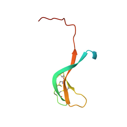
Tick evasins (EVAs) bind either CC- or CXC-chemokines by a poorly understood promiscuous or "one-to-many" mechanism to neutralize inflammation. Because EVAs potently inhibit inflammation in many preclinical models, highlighting their potential as biological therapeutics for inflammatory diseases, we sought to further unravel the CXC-chemokine-EVA interactions. Using yeast surface display, we identified and characterized 27 novel CXC-chemokine-binding evasins homologous to EVA3 and defined two functional classes. The first, which included EVA3, exclusively bound ELR + CXC-chemokines, whereas the second class bound both ELR + and ELR - CXC-chemokines, in several cases including C X C-motif chemokine ligand 10 (CXCL10) but, surprisingly, not CXCL8. The X-ray crystal structure of EVA3 at a resolution of 1.79 Å revealed a single antiparallel β-sheet with six conserved cysteine residues forming a disulfide-bonded knottin scaffold that creates a contiguous solvent-accessible surface. Swapping analyses identified distinct knottin scaffold segments necessary for different CXC-chemokine-binding activities, implying that differential ligand positioning, at least in part, plays a role in promiscuous binding. Swapping segments also transferred chemokine-binding activity, resulting in a hybrid EVA with dual CXCL10- and CXCL8-binding activities. The solvent-accessible surfaces of the knottin scaffold segments have distinctive shape and charge, which we suggest drives chemokine-binding specificity. These studies provide structural and mechanistic insight into how CXC-chemokine-binding tick EVAs achieve class specificity but also engage in promiscuous binding.
Organizational Affiliation:
Radcliffe Department of Medicine Division of Cardiovascular Medicine, University of Oxford, Oxford OX3 7BN, United Kingdom.