6HHO

Crystal structure of RIP1 kinase in complex with GSK547


Experimental Data Snapshot

  • Method: X-RAY DIFFRACTION
  • Resolution: 3.49 Å
  • R-Value Free: 0.227 
  • R-Value Work: 0.208 
  • R-Value Observed: 0.209 

wwPDB Validation   3D Report Full Report


Ligand Structure Quality Assessment 


This is version 1.1 of the entry. See complete history


Literature

RIP1 Kinase Drives Macrophage-Mediated Adaptive Immune Tolerance in Pancreatic Cancer.

Wang, W.Marinis, J.M.Beal, A.M.Savadkar, S.Wu, Y.Khan, M.Taunk, P.S.Wu, N.Su, W.Wu, J.Ahsan, A.Kurz, E.Chen, T.Yaboh, I.Li, F.Gutierrez, J.Diskin, B.Hundeyin, M.Reilly, M.Lich, J.D.Harris, P.A.Mahajan, M.K.Thorpe, J.H.Nassau, P.Mosley, J.E.Leinwand, J.Kochen Rossi, J.A.Mishra, A.Aykut, B.Glacken, M.Ochi, A.Verma, N.Kim, J.I.Vasudevaraja, V.Adeegbe, D.Almonte, C.Bagdatlioglu, E.Cohen, D.J.Wong, K.K.Bertin, J.Miller, G.

(2018) Cancer Cell 34: 757-774.e7

  • DOI: https://doi.org/10.1016/j.ccell.2018.10.006
  • Primary Citation of Related Structures:  
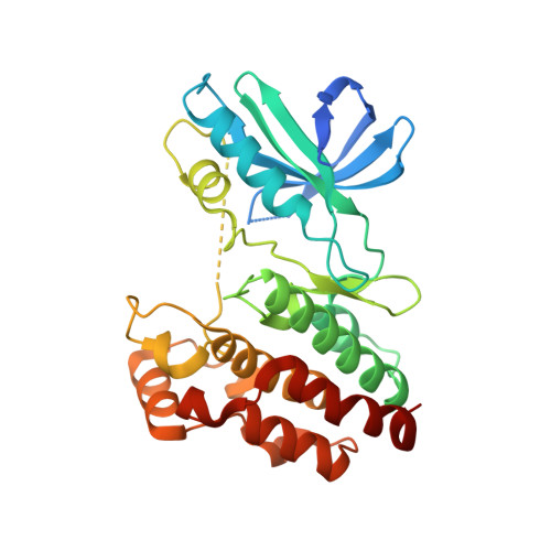
    6HHO

  • PubMed Abstract: 

    Pancreatic ductal adenocarcinoma (PDA) is characterized by immune tolerance and immunotherapeutic resistance. We discovered upregulation of receptor-interacting serine/threonine protein kinase 1 (RIP1) in tumor-associated macrophages (TAMs) in PDA. To study its role in oncogenic progression, we developed a selective small-molecule RIP1 inhibitor with high in vivo exposure. Targeting RIP1 reprogrammed TAMs toward an MHCII hi TNFα + IFNγ + immunogenic phenotype in a STAT1-dependent manner. RIP1 inhibition in TAMs resulted in cytotoxic T cell activation and T helper cell differentiation toward a mixed Th1/Th17 phenotype, leading to tumor immunity in mice and in organotypic models of human PDA. Targeting RIP1 synergized with PD1-and inducible co-stimulator-based immunotherapies. Tumor-promoting effects of RIP1 were independent of its co-association with RIP3. Collectively, our work describes RIP1 as a checkpoint kinase governing tumor immunity.


  • Organizational Affiliation

    S. Arthur Localio Laboratory, Department of Surgery, New York University School of Medicine, 435 East 30th Street, 4th Floor, New York, NY 10016, USA.


Macromolecules
Find similar proteins by:  (by identity cutoff)  |  3D Structure
Entity ID: 1
MoleculeChains Sequence LengthOrganismDetailsImage
Receptor-interacting serine/threonine-protein kinase 1
A, B
303Homo sapiensMutation(s): 4 
Gene Names: RIPK1RIPRIP1
EC: 2.7.11.1
UniProt & NIH Common Fund Data Resources
Find proteins for Q13546 (Homo sapiens)
Explore Q13546 
Go to UniProtKB:  Q13546
PHAROS:  Q13546
GTEx:  ENSG00000137275 
Entity Groups  
Sequence Clusters30% Identity50% Identity70% Identity90% Identity95% Identity100% Identity
UniProt GroupQ13546
Sequence Annotations
Expand
  • Reference Sequence
Small Molecules
Ligands 1 Unique
IDChains Name / Formula / InChI Key2D Diagram3D Interactions
G4W (Subject of Investigation/LOI)
Query on G4W

Download Ideal Coordinates CCD File 
C [auth A],
D [auth B]
6-[4-[(5~{S})-5-[3,5-bis(fluoranyl)phenyl]pyrazolidin-1-yl]carbonylpiperidin-1-yl]pyrimidine-4-carbonitrile
C20 H20 F2 N6 O
PZXPEVAQKNYNRX-SFHVURJKSA-N
Experimental Data & Validation

Experimental Data

  • Method: X-RAY DIFFRACTION
  • Resolution: 3.49 Å
  • R-Value Free: 0.227 
  • R-Value Work: 0.208 
  • R-Value Observed: 0.209 
  • Space Group: P 21 21 21
Unit Cell:
Length ( Å )Angle ( ˚ )
a = 102.649α = 90
b = 130.37β = 90
c = 50.839γ = 90
Software Package:
Software NamePurpose
MOSFLMdata reduction
Aimlessdata scaling
BUSTERrefinement
PDB_EXTRACTdata extraction

Structure Validation

View Full Validation Report


Ligand Structure Quality Assessment 


Entry History 

Deposition Data

Revision History  (Full details and data files)

  • Version 1.0: 2018-12-12
    Type: Initial release
  • Version 1.1: 2024-01-17
    Changes: Data collection, Database references, Refinement description