6H9F

Structure of glutamate mutase reconstituted with bishomo-coenzyme B12


Experimental Data Snapshot

  • Method: X-RAY DIFFRACTION
  • Resolution: 2.10 Å
  • R-Value Free: 0.203 
  • R-Value Work: 0.158 
  • R-Value Observed: 0.160 

wwPDB Validation   3D Report Full Report


Ligand Structure Quality Assessment 


This is version 1.2 of the entry. See complete history


Literature

Structure-Based Demystification of Radical Catalysis by a Coenzyme B 12 Dependent Enzyme-Crystallographic Study of Glutamate Mutase with Cofactor Homologues.

Gruber, K.Csitkovits, V.Lyskowski, A.Kratky, C.Krautler, B.

(2022) Angew Chem Int Ed Engl 61: e202208295-e202208295

  • DOI: https://doi.org/10.1002/anie.202208295
  • Primary Citation of Related Structures:  
    6H9E, 6H9F

  • PubMed Abstract: 

    Catalysis by radical enzymes dependent on coenzyme B 12 (AdoCbl) relies on the reactive primary 5'-deoxy-5'adenosyl radical, which originates from reversible Co-C bond homolysis of AdoCbl. This bond homolysis is accelerated roughly 10 12 -fold upon binding the enzyme substrate. The structural basis for this activation is still strikingly enigmatic. As revealed here, a displaced firm adenosine binding cavity in substrate-loaded glutamate mutase (GM) causes a structural misfit for intact AdoCbl that is relieved by the homolytic Co-C bond cleavage. Strategically interacting adjacent adenosine- and substrate-binding protein cavities provide a tight caged radical reaction space, controlling the entire radical path. The GM active site is perfectly structured for promoting radical catalysis, including "negative catalysis", a paradigm for AdoCbl-dependent mutases.


  • Organizational Affiliation

    Institute of Molecular Biosciences, University of Graz, Humboldtstraße 50, 8010, Graz, Austria.


Macromolecules
Find similar proteins by:  (by identity cutoff)  |  3D Structure
Entity ID: 1
MoleculeChains Sequence LengthOrganismDetailsImage
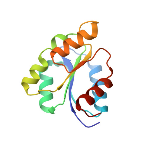
Glutamate mutase sigma subunit
A, C
137Clostridium cochleariumMutation(s): 0 
Gene Names: glmS
EC: 5.4.99.1
UniProt
Find proteins for P80078 (Clostridium cochlearium)
Explore P80078 
Go to UniProtKB:  P80078
Entity Groups  
Sequence Clusters30% Identity50% Identity70% Identity90% Identity95% Identity100% Identity
UniProt GroupP80078
Sequence Annotations
Expand
  • Reference Sequence
Find similar proteins by:  (by identity cutoff)  |  3D Structure
Entity ID: 2
MoleculeChains Sequence LengthOrganismDetailsImage
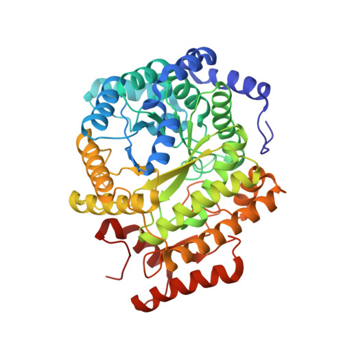
Glutamate mutase epsilon subunit
B, D
483Clostridium cochleariumMutation(s): 0 
Gene Names: glmE
EC: 5.4.99.1
UniProt
Find proteins for P80077 (Clostridium cochlearium)
Explore P80077 
Go to UniProtKB:  P80077
Entity Groups  
Sequence Clusters30% Identity50% Identity70% Identity90% Identity95% Identity100% Identity
UniProt GroupP80077
Sequence Annotations
Expand
  • Reference Sequence
Experimental Data & Validation

Experimental Data

  • Method: X-RAY DIFFRACTION
  • Resolution: 2.10 Å
  • R-Value Free: 0.203 
  • R-Value Work: 0.158 
  • R-Value Observed: 0.160 
  • Space Group: P 1 21 1
Unit Cell:
Length ( Å )Angle ( ˚ )
a = 63.749α = 90
b = 112.286β = 95.712
c = 108.01γ = 90
Software Package:
Software NamePurpose
PHENIXrefinement
MOSFLMdata reduction
Aimlessdata scaling
PHENIXphasing

Structure Validation

View Full Validation Report


Ligand Structure Quality Assessment 


Entry History & Funding Information

Deposition Data


Funding OrganizationLocationGrant Number
Austrian Science FundAustria--

Revision History  (Full details and data files)

  • Version 1.0: 2019-08-14
    Type: Initial release
  • Version 1.1: 2022-10-26
    Changes: Database references, Derived calculations
  • Version 1.2: 2024-01-31
    Changes: Data collection, Refinement description