6H0R

X-ray structure of SRS2 fragment of Rgs4 3' UTR


Experimental Data Snapshot

  • Method: X-RAY DIFFRACTION
  • Resolution: 1.73 Å
  • R-Value Free: 0.237 
  • R-Value Work: 0.185 
  • R-Value Observed: 0.187 

wwPDB Validation   3D Report Full Report


This is version 1.1 of the entry. See complete history


Literature

Staufen2-mediated RNA recognition and localization requires combinatorial action of multiple domains.

Heber, S.Gaspar, I.Tants, J.N.Gunther, J.Moya, S.M.F.Janowski, R.Ephrussi, A.Sattler, M.Niessing, D.

(2019) Nat Commun 10: 1659-1659

  • DOI: https://doi.org/10.1038/s41467-019-09655-3
  • Primary Citation of Related Structures:  
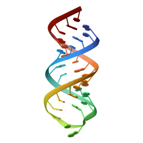
    6H0R

  • PubMed Abstract: 

    Throughout metazoans, Staufen (Stau) proteins are core factors of mRNA localization particles. They consist of three to four double-stranded RNA binding domains (dsRBDs) and a C-terminal dsRBD-like domain. Mouse Staufen2 (mStau2)-like Drosophila Stau (dmStau) contains four dsRBDs. Existing data suggest that only dsRBDs 3-4 are necessary and sufficient for mRNA binding. Here, we show that dsRBDs 1 and 2 of mStau2 bind RNA with similar affinities and kinetics as dsRBDs 3 and 4. While RNA binding by these tandem domains is transient, all four dsRBDs recognize their target RNAs with high stability. Rescue experiments in Drosophila oocytes demonstrate that mStau2 partially rescues dmStau-dependent mRNA localization. In contrast, a rescue with mStau2 bearing RNA-binding mutations in dsRBD1-2 fails, confirming the physiological relevance of our findings. In summary, our data show that the dsRBDs 1-2 play essential roles in the mRNA recognition and function of Stau-family proteins of different species.


  • Organizational Affiliation

    Institute of Pharmaceutical Biotechnology, 89081 Ulm University, Ulm, Germany.


Macromolecules
Find similar nucleic acids by:  (by identity cutoff)  |  3D Structure
Entity ID: 1
MoleculeChains LengthOrganismImage
SRS2 fragment of Rgs4 3' UTR, RNA (31-MER)
A, B
31Rattus norvegicus
Sequence Annotations
Expand
  • Reference Sequence
Small Molecules
Ligands 2 Unique
IDChains Name / Formula / InChI Key2D Diagram3D Interactions
BA
Query on BA

Download Ideal Coordinates CCD File 
C [auth A]
D [auth A]
E [auth A]
F [auth A]
G [auth A]
C [auth A],
D [auth A],
E [auth A],
F [auth A],
G [auth A],
H [auth A],
J [auth B],
K [auth B],
L [auth B],
M [auth B],
N [auth B],
O [auth B]
BARIUM ION
Ba
XDFCIPNJCBUZJN-UHFFFAOYSA-N
MG
Query on MG

Download Ideal Coordinates CCD File 
I [auth A]MAGNESIUM ION
Mg
JLVVSXFLKOJNIY-UHFFFAOYSA-N
Experimental Data & Validation

Experimental Data

  • Method: X-RAY DIFFRACTION
  • Resolution: 1.73 Å
  • R-Value Free: 0.237 
  • R-Value Work: 0.185 
  • R-Value Observed: 0.187 
  • Space Group: C 1 2 1
Unit Cell:
Length ( Å )Angle ( ˚ )
a = 114.02α = 90
b = 32.39β = 103.47
c = 46.37γ = 90
Software Package:
Software NamePurpose
PHENIXrefinement
XDSdata reduction
SCALAdata scaling
Auto-Rickshawphasing

Structure Validation

View Full Validation Report


Entry History 

Deposition Data

Revision History  (Full details and data files)

  • Version 1.0: 2019-04-17
    Type: Initial release
  • Version 1.1: 2019-04-24
    Changes: Data collection, Database references