6GY8

Crystal structure of XaxA from Xenorhabdus nematophila


Experimental Data Snapshot

  • Method: X-RAY DIFFRACTION
  • Resolution: 2.50 Å
  • R-Value Free: 0.286 
  • R-Value Work: 0.238 
  • R-Value Observed: 0.241 

wwPDB Validation   3D Report Full Report


This is version 1.0 of the entry. See complete history


Literature

Membrane insertion of alpha-xenorhabdolysin in near-atomic detail.

Schubert, E.Vetter, I.R.Prumbaum, D.Penczek, P.A.Raunser, S.

(2018) Elife 7

  • DOI: https://doi.org/10.7554/eLife.38017
  • Primary Citation of Related Structures:  
    6GY6, 6GY7, 6GY8

  • PubMed Abstract: 

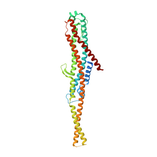
    α-Xenorhabdolysins (Xax) are α-pore-forming toxins (α-PFT) that form 1-1.3 MDa large pore complexes to perforate the host cell membrane. PFTs are used by a variety of bacterial pathogens to attack host cells. Due to the lack of structural information, the molecular mechanism of action of Xax toxins is poorly understood. Here, we report the cryo-EM structure of the XaxAB pore complex from Xenorhabdus nematophila and the crystal structures of the soluble monomers of XaxA and XaxB. The structures reveal that XaxA and XaxB are built similarly and appear as heterodimers in the 12-15 subunits containing pore, classifying XaxAB as bi-component α-PFT. Major conformational changes in XaxB, including the swinging out of an amphipathic helix are responsible for membrane insertion. XaxA acts as an activator and stabilizer for XaxB that forms the actual transmembrane pore. Based on our results, we propose a novel structural model for the mechanism of Xax intoxication.


  • Organizational Affiliation

    Department of Structural Biochemistry, Max Planck Institute of Molecular Physiology, Dortmund, Germany.


Macromolecules
Find similar proteins by:  (by identity cutoff)  |  3D Structure
Entity ID: 1
MoleculeChains Sequence LengthOrganismDetailsImage
XaxA
A, B
408Xenorhabdus nematophila ATCC 19061Mutation(s): 0 
Gene Names: xaxAXNC1_3766
Membrane Entity: Yes 
UniProt
Find proteins for D3VB22 (Xenorhabdus nematophila (strain ATCC 19061 / DSM 3370 / CCUG 14189 / LMG 1036 / NCIMB 9965 / AN6))
Explore D3VB22 
Go to UniProtKB:  D3VB22
Entity Groups  
Sequence Clusters30% Identity50% Identity70% Identity90% Identity95% Identity100% Identity
UniProt GroupD3VB22
Sequence Annotations
Expand
  • Reference Sequence
Experimental Data & Validation

Experimental Data

  • Method: X-RAY DIFFRACTION
  • Resolution: 2.50 Å
  • R-Value Free: 0.286 
  • R-Value Work: 0.238 
  • R-Value Observed: 0.241 
  • Space Group: P 21 21 21
Unit Cell:
Length ( Å )Angle ( ˚ )
a = 67.27α = 90
b = 90.83β = 90
c = 153.04γ = 90
Software Package:
Software NamePurpose
PHENIXrefinement
XDSdata reduction
XSCALEdata scaling
HKL2Mapphasing

Structure Validation

View Full Validation Report


Entry History & Funding Information

Deposition Data


Funding OrganizationLocationGrant Number
European Research CouncilGermany615984

Revision History  (Full details and data files)

  • Version 1.0: 2018-07-25
    Type: Initial release