6GRT

Paired immunoglobulin-like receptor B (PirB) or Leukocyte immunoglobulin-like receptor subfamily B member 3 (LILRB3) full extracellular domain


Experimental Data Snapshot

  • Method: X-RAY DIFFRACTION
  • Resolution: 4.50 Å
  • R-Value Free: 0.307 
  • R-Value Work: 0.259 
  • R-Value Observed: 0.261 

wwPDB Validation   3D Report Full Report


Ligand Structure Quality Assessment 


This is version 2.1 of the entry. See complete history


Literature

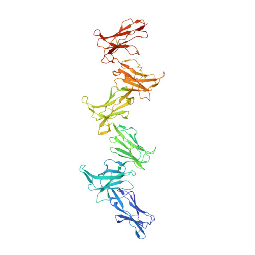
Structure and flexibility of the extracellular region of the PirB receptor.

Vlieg, H.C.Huizinga, E.G.Janssen, B.J.C.

(2019) J Biol Chem 294: 4634-4643

  • DOI: https://doi.org/10.1074/jbc.RA118.004396
  • Primary Citation of Related Structures:  
    6GRQ, 6GRS, 6GRT

  • PubMed Abstract: 

    Murine paired immunoglobulin receptor B (PirB) and its human ortholog leukocyte immunoglobulin-like receptor B2 (LILRB2) are widely expressed inhibitory receptors that interact with a diverse set of extracellular ligands and exert functions ranging from down-regulation of immune responses to inhibition of neuronal growth. However, structural information that could shed light on how PirB interacts with its ligands is lacking. Here, we report crystal structures of the PirB ectodomain; the first full ectodomain structure for a LILR family member, at 3.3-4.5 Å resolution. The structures reveal that PirB's six Ig-like domains are arranged at acute angles, similar to the structures of leukocyte immunoglobulin-like receptor (LILR) and killer-cell immunoglobulin-like receptor (KIR). We observe that this regular arrangement is followed throughout the ectodomain, resulting in an extended zigzag conformation. In two out of the five structures reported here, the repeating zigzag is broken by the first domain that can adopt two alternative orientations. Quantitative binding experiments revealed a 9 μm dissociation constant for PirB-myelin-associated glycoprotein (MAG) ectodomain interactions. Taken together, these structural findings and the observed PirB-MAG interactions are compatible with a model for intercellular signaling in which the PirB extracellular domains, which point away from the cell surface, enable interaction with ligands in trans .


  • Organizational Affiliation

    From Crystal and Structural Chemistry, Bijvoet Center for Biomolecular Research, Department of Chemistry, Faculty of Science, Utrecht University, 3584 CH Utrecht, The Netherlands.


Macromolecules
Find similar proteins by:  (by identity cutoff)  |  3D Structure
Entity ID: 1
MoleculeChains Sequence LengthOrganismDetailsImage
Paired immunoglobulin-like receptor B
A, B
606Mus musculusMutation(s): 0 
Gene Names: PirbLilrb3
UniProt
Find proteins for Q8K4V6 (Mus musculus)
Explore Q8K4V6 
Go to UniProtKB:  Q8K4V6
Entity Groups  
Sequence Clusters30% Identity50% Identity70% Identity90% Identity95% Identity100% Identity
UniProt GroupQ8K4V6
Sequence Annotations
Expand
  • Reference Sequence
Oligosaccharides

Help

Entity ID: 2
MoleculeChains Length2D Diagram Glycosylation3D Interactions
beta-D-mannopyranose-(1-4)-2-acetamido-2-deoxy-beta-D-glucopyranose-(1-4)-2-acetamido-2-deoxy-beta-D-glucopyranose
C
3N-Glycosylation
Glycosylation Resources
GlyTouCan:  G15407YE
GlyCosmos:  G15407YE
GlyGen:  G15407YE
Entity ID: 3
MoleculeChains Length2D Diagram Glycosylation3D Interactions
alpha-D-mannopyranose-(1-3)-[alpha-D-mannopyranose-(1-6)]beta-D-mannopyranose-(1-4)-2-acetamido-2-deoxy-beta-D-glucopyranose-(1-4)-2-acetamido-2-deoxy-beta-D-glucopyranose
D
5N-Glycosylation
Glycosylation Resources
GlyTouCan:  G22768VO
GlyCosmos:  G22768VO
GlyGen:  G22768VO
Small Molecules
Ligands 1 Unique
IDChains Name / Formula / InChI Key2D Diagram3D Interactions
NAG
Query on NAG

Download Ideal Coordinates CCD File 
E [auth A],
F [auth B]
2-acetamido-2-deoxy-beta-D-glucopyranose
C8 H15 N O6
OVRNDRQMDRJTHS-FMDGEEDCSA-N
Experimental Data & Validation

Experimental Data

  • Method: X-RAY DIFFRACTION
  • Resolution: 4.50 Å
  • R-Value Free: 0.307 
  • R-Value Work: 0.259 
  • R-Value Observed: 0.261 
  • Space Group: P 1 21 1
Unit Cell:
Length ( Å )Angle ( ˚ )
a = 67.254α = 90
b = 127.103β = 103.43
c = 144.08γ = 90
Software Package:
Software NamePurpose
PHENIXrefinement
iMOSFLMdata reduction
Aimlessdata scaling
PHASERphasing

Structure Validation

View Full Validation Report


Ligand Structure Quality Assessment 


Entry History & Funding Information

Deposition Data


Funding OrganizationLocationGrant Number
Netherlands Organisation for Scientific ResearchNetherlands723.012.002

Revision History  (Full details and data files)

  • Version 1.0: 2019-02-06
    Type: Initial release
  • Version 1.1: 2019-04-03
    Changes: Data collection, Database references
  • Version 2.0: 2020-07-29
    Type: Remediation
    Reason: Carbohydrate remediation
    Changes: Atomic model, Data collection, Derived calculations, Structure summary
  • Version 2.1: 2024-01-17
    Changes: Data collection, Database references, Derived calculations, Refinement description, Structure summary