6GBR

Crystal Structure of the oligomerization domain of VP35 from Reston virus, mercury derivative


Experimental Data Snapshot

  • Method: X-RAY DIFFRACTION
  • Resolution: 3.15 Å
  • R-Value Free: 0.311 
  • R-Value Work: 0.207 
  • R-Value Observed: 0.212 

wwPDB Validation   3D Report Full Report


Ligand Structure Quality Assessment 


This is version 1.2 of the entry. See complete history


Literature

Structures of Ebola and Reston Virus VP35 Oligomerization Domains and Comparative Biophysical Characterization in All Ebolavirus Species.

Zinzula, L.Nagy, I.Orsini, M.Weyher-Stingl, E.Bracher, A.Baumeister, W.

(2019) Structure 27: 39-54.e6

  • DOI: https://doi.org/10.1016/j.str.2018.09.009
  • Primary Citation of Related Structures:  
    6GBO, 6GBP, 6GBQ, 6GBR

  • PubMed Abstract: 

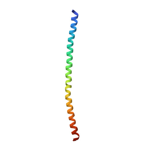
    The multifunctional virion protein 35 (VP35) of ebolaviruses is a critical determinant of virulence and pathogenesis indispensable for viral replication and host innate immune evasion. Essential for VP35 function is homo-oligomerization via a coiled-coil motif. Here we report crystal structures of VP35 oligomerization domains from the prototypic Ebola virus (EBOV) and the non-pathogenic Reston virus (RESTV), together with a comparative biophysical characterization of the domains from all known species of the Ebolavirus genus. EBOV and RESTV VP35 oligomerization domains form bipartite parallel helix bundles with a canonical coiled coil in the N-terminal half and increased plasticity in the highly conserved C-terminal half. The domain assembles into trimers and tetramers in EBOV, whereas it exclusively forms tetramers in all other ebolavirus species. Substitution of coiled-coil leucine residues critical for immune antagonism leads to aberrant oligomerization. A conserved arginine involved in inter-chain salt bridges stabilizes the VP35 oligomerization domain and modulates between coiled-coil oligomeric states.


  • Organizational Affiliation

    The Max-Planck Institute of Biochemistry, Department of Molecular Structural Biology, Am Klopferspitz 18, 82152 Martinsried, Germany.


Macromolecules
Find similar proteins by:  (by identity cutoff)  |  3D Structure
Entity ID: 1
MoleculeChains Sequence LengthOrganismDetailsImage
Polymerase cofactor VP35
A, B, C, D
71Reston ebolavirusMutation(s): 0 
Gene Names: VP35REBOVgp2
UniProt
Find proteins for Q8JPY0 (Reston ebolavirus (strain Reston-89))
Explore Q8JPY0 
Go to UniProtKB:  Q8JPY0
Entity Groups  
Sequence Clusters30% Identity50% Identity70% Identity90% Identity95% Identity100% Identity
UniProt GroupQ8JPY0
Sequence Annotations
Expand
  • Reference Sequence
Small Molecules
Experimental Data & Validation

Experimental Data

  • Method: X-RAY DIFFRACTION
  • Resolution: 3.15 Å
  • R-Value Free: 0.311 
  • R-Value Work: 0.207 
  • R-Value Observed: 0.212 
  • Space Group: P 21 21 21
Unit Cell:
Length ( Å )Angle ( ˚ )
a = 35.933α = 90
b = 91.901β = 90
c = 104.193γ = 90
Software Package:
Software NamePurpose
XDSdata reduction
Aimlessdata scaling
SHELXDEphasing
PHENIXrefinement
PDB_EXTRACTdata extraction

Structure Validation

View Full Validation Report


Ligand Structure Quality Assessment 


Entry History 

Deposition Data

Revision History  (Full details and data files)

  • Version 1.0: 2018-10-10
    Type: Initial release
  • Version 1.1: 2018-12-12
    Changes: Data collection, Database references, Structure summary
  • Version 1.2: 2019-01-16
    Changes: Data collection, Database references