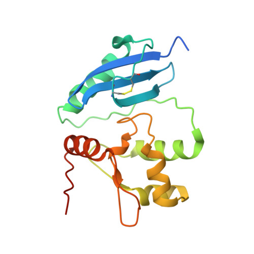
Crystal structure of a thermophilic O6-alkylguanine-DNA alkyltransferase-derived self-labeling protein-tag in covalent complex with a fluorescent probe.
Rossi, F., Morrone, C., Massarotti, A., Ferraris, D.M., Valenti, A., Perugino, G., Miggiano, R.(2018) Biochem Biophys Res Commun 500: 698-703
- PubMed: 29684348
- DOI: https://doi.org/10.1016/j.bbrc.2018.04.139
- Primary Citation of Related Structures:
6GA0 - PubMed Abstract:
The self-labeling protein tags are robust and versatile tools for studying different molecular aspects of cell biology. In order to be suitable for a wide spectrum of experimental conditions, it is mandatory that these systems are stable after the fluorescent labeling reaction and do not alter the properties of the fusion partner. SsOGT-H 5 is an engineered variant alkylguanine-DNA-alkyl-transferase (OGT) of the hyperthermophilic archaeon Sulfolobus solfataricus, and it represents an alternative solution to the SNAP-tag ® technology under harsh reaction conditions. Here we present the crystal structure of SsOGT-H 5 in complex with the fluorescent probe SNAP-Vista Green ® (SsOGT-H 5 -SVG) that reveals the conformation adopted by the protein upon the trans-alkylation reaction with the substrate, which is observed covalently bound to the catalytic cysteine residue. Moreover, we identify the amino acids that contribute to both the overall protein stability in the post-reaction state and the coordination of the fluorescent moiety stretching-out from the protein active site. We gained new insights in the conformational changes possibly occurring to the OGT proteins upon reaction with modified guanine base bearing bulky adducts; indeed, our structural analysis reveals an unprecedented conformation of the active site loop that is likely to trigger protein destabilization and consequent degradation. Interestingly, the SVG moiety plays a key role in restoring the interaction between the N- and C-terminal domains of the protein that is lost following the new conformation adopted by the active site loop in the SsOGT-H 5 -SVG structure. Molecular dynamics simulations provide further information into the dynamics of SsOGT-H 5 -SVG structure, highlighting the role of the fluorescent ligand in keeping the protein stable after the trans-alkylation reaction.
Organizational Affiliation:
DSF-Dipartimento di Scienze del Farmaco, University of Piemonte Orientale, Via Bovio 6, 28100 Novara, Italy. Electronic address: franca.rossi@uniupo.it.