


Structural basis of inactivation of Enterococcus faecium penicillin binding protein 5 by ceftobiprole.
Sauvage, E., El Gachi, M., Kerff, F., Herman, R., Verlaine, O., Amoroso, A., Page, M.G.P., Joris, B., Charlier, P.To be published.
Experimental Data Snapshot
Entity ID: 1 | |||||
|---|---|---|---|---|---|
| Molecule | Chains | Sequence Length | Organism | Details | Image |
| Low affinity penicillin-binding protein 5 (PBP5) | 649 | Enterococcus faecium | Mutation(s): 0 Gene Names: pbp5 | 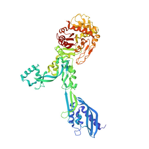 | |
UniProt | |||||
Find proteins for Q47759 (Enterococcus faecium) Explore Q47759 Go to UniProtKB: Q47759 | |||||
Entity Groups | |||||
| Sequence Clusters | 30% Identity50% Identity70% Identity90% Identity95% Identity100% Identity | ||||
| UniProt Group | Q47759 | ||||
Sequence AnnotationsExpand | |||||
| |||||
| Ligands 2 Unique | |||||
|---|---|---|---|---|---|
| ID | Chains | Name / Formula / InChI Key | 2D Diagram | 3D Interactions | |
| RB6 Query on RB6 | D [auth A], F [auth B], H [auth C] | (2R)-2-[(1R)-1-{[(2Z)-2-(5-amino-1,2,4-thiadiazol-3-yl)-2-(hydroxyimino)acetyl]amino}-2-oxoethyl]-5-({2-oxo-1-[(3R)-pyr
rolidin-3-yl]-2,5-dihydro-1H-pyrrol-3-yl}methyl)-3,6-dihydro-2H-1,3-thiazine-4-carboxylic acid C20 H24 N8 O6 S2 MYAXGJQBOYOEHQ-SWBIIUODSA-N |  | ||
| SO4 Query on SO4 | E [auth A], G [auth B], I [auth C] | SULFATE ION O4 S QAOWNCQODCNURD-UHFFFAOYSA-L |  | ||
| Length ( Å ) | Angle ( ˚ ) |
|---|---|
| a = 79.194 | α = 90 |
| b = 128.736 | β = 94.04 |
| c = 238.479 | γ = 90 |
| Software Name | Purpose |
|---|---|
| PHENIX | refinement |
| XDS | data reduction |
| XSCALE | data scaling |
| AMoRE | phasing |
RCSB PDB (citation) is hosted by
RCSB PDB is a member of the

