6G6L

The crystal structures of Human MYC:MAX bHLHZip complex


Experimental Data Snapshot

  • Method: X-RAY DIFFRACTION
  • Resolution: 2.20 Å
  • R-Value Free: 0.268 
  • R-Value Work: 0.215 
  • R-Value Observed: 0.217 

wwPDB Validation   3D Report Full Report


This is version 1.3 of the entry. See complete history


Literature

Crystal Structures and Nuclear Magnetic Resonance Studies of the Apo Form of the c-MYC:MAX bHLHZip Complex Reveal a Helical Basic Region in the Absence of DNA.

Sammak, S.Hamdani, N.Gorrec, F.Allen, M.D.Freund, S.M.V.Bycroft, M.Zinzalla, G.

(2019) Biochemistry 58: 3144-3154

  • DOI: https://doi.org/10.1021/acs.biochem.9b00296
  • Primary Citation of Related Structures:  
    6G6J, 6G6K, 6G6L

  • PubMed Abstract: 

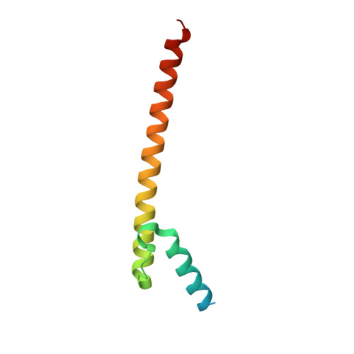
    The c-MYC transcription factor is a master regulator of cell growth and proliferation and is an established target for cancer therapy. This basic helix-loop-helix Zip protein forms a heterodimer with its obligatory partner MAX, which binds to DNA via the basic region. Considerable research efforts are focused on targeting the heterodimerization interface and the interaction of the complex with DNA. The only available crystal structure is that of a c-MYC:MAX complex artificially tethered by an engineered disulfide linker and prebound to DNA. We have carried out a detailed structural analysis of the apo form of the c-MYC:MAX complex, with no artificial linker, both in solution using nuclear magnetic resonance (NMR) spectroscopy and by X-ray crystallography. We have obtained crystal structures in three different crystal forms, with resolutions between 1.35 and 2.2 Å, that show extensive helical structure in the basic region. Determination of the α-helical propensity using NMR chemical shift analysis shows that the basic region of c-MYC and, to a lesser extent, that of MAX populate helical conformations. We have also assigned the NMR spectra of the c-MYC basic helix-loop-helix Zip motif in the absence of MAX and showed that the basic region has an intrinsic helical propensity even in the absence of its dimerization partner. The presence of helical structure in the basic regions in the absence of DNA suggests that the molecular recognition occurs via a conformational selection rather than an induced fit. Our work provides both insight into the mechanism of DNA binding and structural information to aid in the development of MYC inhibitors.


  • Organizational Affiliation

    Microbiology, Tumor and Cell Biology (MTC) , Karolinska Institutet , Solnavägen 9 , 171 65 Stockholm , Sweden.


Macromolecules
Find similar proteins by:  (by identity cutoff)  |  3D Structure
Entity ID: 1
MoleculeChains Sequence LengthOrganismDetailsImage
Myc proto-oncogene protein
A, C, E, G
94Homo sapiensMutation(s): 0 
Gene Names: MYCBHLHE39
UniProt & NIH Common Fund Data Resources
Find proteins for P01106 (Homo sapiens)
Explore P01106 
Go to UniProtKB:  P01106
PHAROS:  P01106
GTEx:  ENSG00000136997 
Entity Groups  
Sequence Clusters30% Identity50% Identity70% Identity90% Identity95% Identity100% Identity
UniProt GroupP01106
Sequence Annotations
Expand
  • Reference Sequence
Find similar proteins by:  (by identity cutoff)  |  3D Structure
Entity ID: 2
MoleculeChains Sequence LengthOrganismDetailsImage
Protein max
B, D, F, H
83Homo sapiensMutation(s): 0 
Gene Names: MAXBHLHD4
UniProt & NIH Common Fund Data Resources
Find proteins for P61244 (Homo sapiens)
Explore P61244 
Go to UniProtKB:  P61244
PHAROS:  P61244
GTEx:  ENSG00000125952 
Entity Groups  
Sequence Clusters30% Identity50% Identity70% Identity90% Identity95% Identity100% Identity
UniProt GroupP61244
Sequence Annotations
Expand
  • Reference Sequence
Small Molecules
Ligands 1 Unique
IDChains Name / Formula / InChI Key2D Diagram3D Interactions
SO4
Query on SO4

Download Ideal Coordinates CCD File 
I [auth A]
J [auth A]
K [auth B]
L [auth B]
M [auth C]
I [auth A],
J [auth A],
K [auth B],
L [auth B],
M [auth C],
N [auth D],
O [auth D],
P [auth E],
Q [auth F],
R [auth F],
S [auth G],
T [auth G],
U [auth H],
V [auth H]
SULFATE ION
O4 S
QAOWNCQODCNURD-UHFFFAOYSA-L
Experimental Data & Validation

Experimental Data

  • Method: X-RAY DIFFRACTION
  • Resolution: 2.20 Å
  • R-Value Free: 0.268 
  • R-Value Work: 0.215 
  • R-Value Observed: 0.217 
  • Space Group: P 1
Unit Cell:
Length ( Å )Angle ( ˚ )
a = 48.68α = 107.12
b = 74.33β = 107.67
c = 80.06γ = 90.05
Software Package:
Software NamePurpose
PHENIXrefinement
iMOSFLMdata reduction
SCALAdata scaling
PHASERphasing

Structure Validation

View Full Validation Report


Entry History 

Deposition Data

Revision History  (Full details and data files)

  • Version 1.0: 2019-04-10
    Type: Initial release
  • Version 1.1: 2019-07-24
    Changes: Data collection, Database references
  • Version 1.2: 2019-07-31
    Changes: Data collection, Database references
  • Version 1.3: 2023-04-05
    Changes: Database references, Derived calculations, Experimental preparation