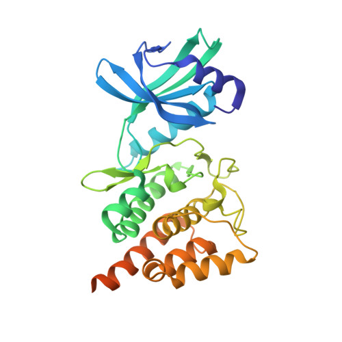
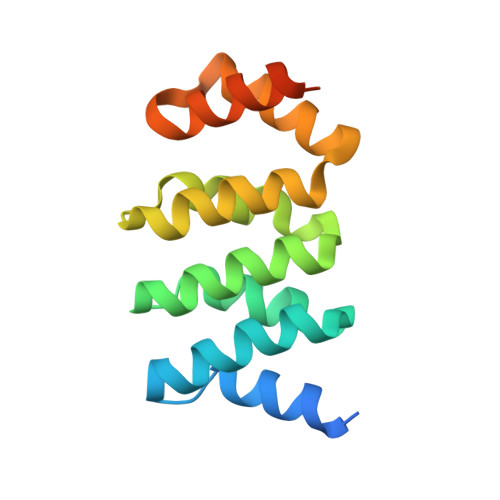
Structural Analysis of the Hanks-Type Protein Kinase YabT FromBacillus subtilisProvides New Insights in its DNA-Dependent Activation.
Shi, L., Cavagnino, A., Rabefiraisana, J.L., Lazar, N., Li de la Sierra-Gallay, I., Ochsenbein, F., Valerio-Lepiniec, M., Urvoas, A., Minard, P., Mijakovic, I., Nessler, S.(2018) Front Microbiol 9: 3014-3014
- PubMed: 30671027
- DOI: https://doi.org/10.3389/fmicb.2018.03014
- Primary Citation of Related Structures:
6G4J - PubMed Abstract:
YabT is a serine/threonine kinase of the Hanks family from Bacillus subtilis , which lacks the canonical extracellular signal receptor domain but is anchored to the membrane through a C-terminal transmembrane helix. A previous study demonstrated that a basic juxtamembrane region corresponds to a DNA-binding motif essential for the activation of YabT trans-autophosphorylation. YabT is expressed during spore development and localizes to the asymmetric septum where it specifically phosphorylates essential proteins involved in genome maintenance, such as RecA, SsbA, and YabA. YabT has also been shown to phosphorylate proteins involved in protein synthesis, such as AbrB and Ef-Tu, suggesting a possible regulatory role in the progressive metabolic quiescence of the forespore. Finally, cross phosphorylations with other protein kinases implicate YabT in the regulation of numerous other cellular processes. Using an artificial protein scaffold as crystallization helper, we determined the first crystal structure of this DNA-dependent bacterial protein kinase. This allowed us to trap the active conformation of the kinase domain of YabT. Using NMR, we showed that the basic juxtamembrane region of YabT is disordered in the absence of DNA in solution, just like it is in the crystal, and that it is stabilized upon DNA binding. In comparison with its closest structural homolog, the mycobacterial kinase PknB allowed us to discuss the dimerization mode of YabT. Together with phosphorylation assays and DNA-binding experiments, this structural analysis helped us to gain new insights into the regulatory activation mechanism of YabT.
Organizational Affiliation:
Division of Systems and Synthetic Biology, Department of Chemical and Biological Engineering, Chalmers University of Technology, Gothenburg, Sweden.