The molecular basis of protein toxin HicA-dependent binding of the protein antitoxin HicB to DNA.
Winter, A.J., Williams, C., Isupov, M.N., Crocker, H., Gromova, M., Marsh, P., Wilkinson, O.J., Dillingham, M.S., Harmer, N.J., Titball, R.W., Crump, M.P.(2018) J Biol Chem 293: 19429-19440
- PubMed: 30337369
- DOI: https://doi.org/10.1074/jbc.RA118.005173
- Primary Citation of Related Structures:
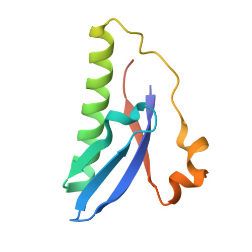
6G1C, 6G1N, 6G26 - PubMed Abstract:
Toxin-antitoxin (TA) systems are present in many bacteria and play important roles in bacterial growth, physiology, and pathogenicity. Those that are best studied are the type II TA systems, in which both toxins and antitoxins are proteins. The HicAB system is one of the prototypic TA systems, found in many bacterial species. Complex interactions between the protein toxin (HicA), the protein antitoxin (HicB), and the DNA upstream of the encoding genes regulate the activity of this system, but few structural details are available about how HicA destabilizes the HicB-DNA complex. Here, we determined the X-ray structures of HicB and the HicAB complex to 1.8 and 2.5 Å resolution, respectively, and characterized their DNA interactions. This revealed that HicB forms a tetramer and HicA and HicB form a heterooctameric complex that involves structural reorganization of the C-terminal (DNA-binding) region of HicB. Our observations indicated that HicA has a profound impact on binding of HicB to DNA sequences upstream of hicAB in a stoichiometric-dependent way. At low ratios of HicA:HicB, there was no effect on DNA binding, but at higher ratios, the affinity for DNA declined cooperatively, driving dissociation of the HicA:HicB:DNA complex. These results reveal the structural mechanisms by which HicA de-represses the HicB-DNA complex.
Organizational Affiliation:
From the School of Chemistry, University of Bristol Cantock's Close, Bristol BS8 1TS, United Kingdom.