6FWU

Crystal structure of human wild type beta-1,4-galactosyltransferase-1 (B4GalT1) in apo-closed dimeric form


Experimental Data Snapshot

  • Method: X-RAY DIFFRACTION
  • Resolution: 2.35 Å
  • R-Value Free: 0.256 
  • R-Value Work: 0.195 
  • R-Value Observed: 0.198 

wwPDB Validation   3D Report Full Report


This is version 1.2 of the entry. See complete history


Literature

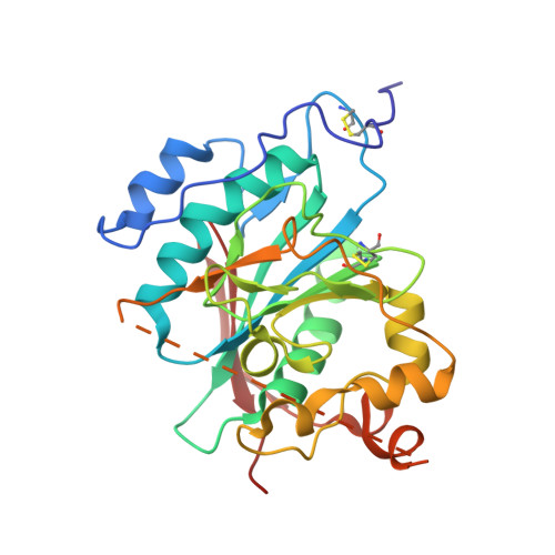
The dimeric structure of wild-type human glycosyltransferase B4GalT1.

Harrus, D.Khoder-Agha, F.Peltoniemi, M.Hassinen, A.Ruddock, L.Kellokumpu, S.Glumoff, T.

(2018) PLoS One 13: e0205571-e0205571

  • DOI: https://doi.org/10.1371/journal.pone.0205571
  • Primary Citation of Related Structures:  
    6FWT, 6FWU

  • PubMed Abstract: 

    Most glycosyltransferases, including B4GalT1 (EC 2.4.1.38), are known to assemble into enzyme homomers and functionally relevant heteromers in vivo. However, it remains unclear why and how these enzymes interact at the molecular/atomic level. Here, we solved the crystal structure of the wild-type human B4GalT1 homodimer. We also show that B4GalT1 exists in a dynamic equilibrium between monomer and dimer, since a purified monomer reappears as a mixture of both and as we obtained crystal forms of the monomer and dimer assemblies in the same crystallization conditions. These two crystal forms revealed the unliganded B4GalT1 in both the open and the closed conformation of the Trp loop and the lid regions, responsible for donor and acceptor substrate binding, respectively. The present structures also show the lid region in full in an open conformation, as well as a new conformation for the GlcNAc acceptor loop (residues 272-288). The physiological relevance of the homodimer in the crystal was validated by targeted mutagenesis studies coupled with FRET assays. These showed that changing key catalytic amino acids impaired homomer formation in vivo. The wild-type human B4GalT1 structure also explains why the variant proteins used for crystallization in earlier studies failed to reveal the homodimers described in this study.


  • Organizational Affiliation

    Faculty of Biochemistry and Molecular Medicine, University of Oulu, Aapistie 7A, Oulu, Finland.


Macromolecules
Find similar proteins by:  (by identity cutoff)  |  3D Structure
Entity ID: 1
MoleculeChains Sequence LengthOrganismDetailsImage
Beta-1,4-galactosyltransferase 1
A, B
273Homo sapiensMutation(s): 0 
Gene Names: B4GALT1GGTB2
EC: 2.4.1 (PDB Primary Data), 2.4.1.22 (PDB Primary Data), 2.4.1.90 (PDB Primary Data), 2.4.1.38 (PDB Primary Data)
UniProt & NIH Common Fund Data Resources
Find proteins for P15291 (Homo sapiens)
Explore P15291 
Go to UniProtKB:  P15291
PHAROS:  P15291
GTEx:  ENSG00000086062 
Entity Groups  
Sequence Clusters30% Identity50% Identity70% Identity90% Identity95% Identity100% Identity
UniProt GroupP15291
Sequence Annotations
Expand
  • Reference Sequence
Experimental Data & Validation

Experimental Data

  • Method: X-RAY DIFFRACTION
  • Resolution: 2.35 Å
  • R-Value Free: 0.256 
  • R-Value Work: 0.195 
  • R-Value Observed: 0.198 
  • Space Group: P 31 2 1
Unit Cell:
Length ( Å )Angle ( ˚ )
a = 60.217α = 90
b = 60.217β = 90
c = 229.368γ = 120
Software Package:
Software NamePurpose
PHENIXrefinement
PDB_EXTRACTdata extraction
XDSdata reduction
XSCALEdata scaling
PHASERphasing

Structure Validation

View Full Validation Report


Entry History & Funding Information

Deposition Data


Funding OrganizationLocationGrant Number
Academy of FinlandFinland285232

Revision History  (Full details and data files)

  • Version 1.0: 2018-10-10
    Type: Initial release
  • Version 1.1: 2018-11-07
    Changes: Data collection, Database references, Structure summary
  • Version 1.2: 2024-01-17
    Changes: Data collection, Database references, Refinement description