


The structure of Schistosoma mansoni Phosphodiesterase 4A
Brown, D.G., Schroeder, S., Gil, C., Munday, J., Koning, H., Siderius, M., Leurs, R.To be published.
Experimental Data Snapshot
wwPDB Validation 3D Report Full Report

Entity ID: 1 | |||||
|---|---|---|---|---|---|
| Molecule | Chains | Sequence Length | Organism | Details | Image |
| Phosphodiesterase | 370 | Schistosoma mansoni | Mutation(s): 0 Gene Names: Smp_134140 EC: 3.1.4 | 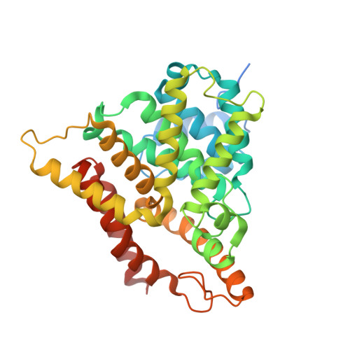 | |
Entity Groups | |||||
| Sequence Clusters | 30% Identity50% Identity70% Identity90% Identity95% Identity100% Identity | ||||
Sequence AnnotationsExpand | |||||
| |||||
| Ligands 2 Unique | |||||
|---|---|---|---|---|---|
| ID | Chains | Name / Formula / InChI Key | 2D Diagram | 3D Interactions | |
| ZN Query on ZN | C [auth A], E [auth B] | ZINC ION Zn PTFCDOFLOPIGGS-UHFFFAOYSA-N |  | ||
| MG Query on MG | D [auth A], F [auth B] | MAGNESIUM ION Mg JLVVSXFLKOJNIY-UHFFFAOYSA-N |  | ||
| Length ( Å ) | Angle ( ˚ ) |
|---|---|
| a = 81.799 | α = 90 |
| b = 81.799 | β = 90 |
| c = 256.127 | γ = 120 |
| Software Name | Purpose |
|---|---|
| REFMAC | refinement |
| XDS | data reduction |
| Aimless | data scaling |
| MxCuBE | data collection |
| PHASER | phasing |
| Funding Organization | Location | Grant Number |
|---|---|---|
| European Union | United Kingdom | EC-FP7-602666-PDE4NPD |
RCSB PDB (citation) is hosted by
RCSB PDB is a member of the

