6F1W

Crystal structure of human Casein Kinase I delta in complex with compound 31a


Experimental Data Snapshot

  • Method: X-RAY DIFFRACTION
  • Resolution: 1.86 Å
  • R-Value Free: 0.206 
  • R-Value Work: 0.181 
  • R-Value Observed: 0.182 

wwPDB Validation   3D Report Full Report


Ligand Structure Quality Assessment 


This is version 1.0 of the entry. See complete history


Literature

Design, Synthesis and Biological Evaluation of Isoxazole-Based CK1 Inhibitors Modified with Chiral Pyrrolidine Scaffolds.

Luxenburger, A.Schmidt, D.Ianes, C.Pichlo, C.Kruger, M.von Drathen, T.Brunstein, E.Gainsford, G.J.Baumann, U.Knippschild, U.Peifer, C.

(2019) Molecules 24

  • DOI: https://doi.org/10.3390/molecules24050873
  • Primary Citation of Related Structures:  
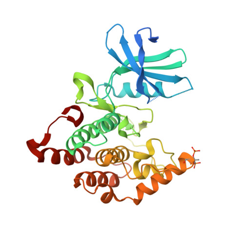
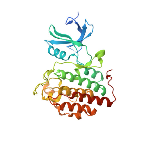
    6F1W, 6F26

  • PubMed Abstract: 

    In this study, we report on the modification of a 3,4-diaryl-isoxazole-based CK1 inhibitor with chiral pyrrolidine scaffolds to develop potent and selective CK1 inhibitors. The pharmacophore of the lead structure was extended towards the ribose pocket of the adenosine triphosphate (ATP) binding site driven by structure-based drug design. For an upscale compatible multigram synthesis of the functionalized pyrrolidine scaffolds, we used a chiral pool synthetic route starting from methionine. Biological evaluation of key compounds in kinase and cellular assays revealed significant effects of the scaffolds towards activity and selectivity, however, the absolute configuration of the chiral moieties only exhibited a limited effect on inhibitory activity. X-ray crystallographic analysis of ligand-CK1δ complexes confirmed the expected binding mode of the 3,4-diaryl-isoxazole inhibitors. Surprisingly, the original compounds underwent spontaneous Pictet-Spengler cyclization with traces of formaldehyde during the co-crystallization process to form highly potent new ligands. Our data suggests chiral "ribose-like" pyrrolidine scaffolds have interesting potential for modifications of pharmacologically active compounds.


  • Organizational Affiliation

    Ferrier Research Institute, Victoria University of Wellington, 69 Gracefield Rd, Lower Hutt 5040, New Zealand. Andreas.Luxenburger@vuw.ac.nz.


Macromolecules
Find similar proteins by:  (by identity cutoff)  |  3D Structure
Entity ID: 1
MoleculeChains Sequence LengthOrganismDetailsImage
Casein kinase I isoform delta314Homo sapiensMutation(s): 0 
Gene Names: CSNK1DHCKID
EC: 2.7.11.1 (PDB Primary Data), 2.7.11.26 (PDB Primary Data)
UniProt & NIH Common Fund Data Resources
Find proteins for P48730 (Homo sapiens)
Explore P48730 
Go to UniProtKB:  P48730
PHAROS:  P48730
GTEx:  ENSG00000141551 
Entity Groups  
Sequence Clusters30% Identity50% Identity70% Identity90% Identity95% Identity100% Identity
UniProt GroupP48730
Sequence Annotations
Expand
  • Reference Sequence
Find similar proteins by:  (by identity cutoff)  |  3D Structure
Entity ID: 2
MoleculeChains Sequence LengthOrganismDetailsImage
Casein kinase I isoform delta314Homo sapiensMutation(s): 0 
Gene Names: CSNK1DHCKID
EC: 2.7.11.1 (PDB Primary Data), 2.7.11.26 (PDB Primary Data)
UniProt & NIH Common Fund Data Resources
Find proteins for P48730 (Homo sapiens)
Explore P48730 
Go to UniProtKB:  P48730
PHAROS:  P48730
GTEx:  ENSG00000141551 
Entity Groups  
Sequence Clusters30% Identity50% Identity70% Identity90% Identity95% Identity100% Identity
UniProt GroupP48730
Sequence Annotations
Expand
  • Reference Sequence
Small Molecules
Ligands 2 Unique
IDChains Name / Formula / InChI Key2D Diagram3D Interactions
CG5
Query on CG5

Download Ideal Coordinates CCD File 
J [auth A],
S [auth B]
(9~{R},10~{R},11~{S})-~{N}-[4-[3-(4-fluorophenyl)-5-propan-2-yl-1,2-oxazol-4-yl]pyridin-2-yl]-4-(4-methoxyphenyl)-10,11-bis(oxidanyl)-1,7-diazatricyclo[7.3.0.0^{3,7}]dodeca-3,5-diene-6-carboxamide
C35 H34 F N5 O5
VRODZCFGQSWGFE-ZLWRCJDJSA-N
SO4
Query on SO4

Download Ideal Coordinates CCD File 
C [auth A]
D [auth A]
E [auth A]
F [auth A]
G [auth A]
C [auth A],
D [auth A],
E [auth A],
F [auth A],
G [auth A],
H [auth A],
I [auth A],
K [auth B],
L [auth B],
M [auth B],
N [auth B],
O [auth B],
P [auth B],
Q [auth B],
R [auth B]
SULFATE ION
O4 S
QAOWNCQODCNURD-UHFFFAOYSA-L
Modified Residues  1 Unique
IDChains TypeFormula2D DiagramParent
TPO
Query on TPO
A
L-PEPTIDE LINKINGC4 H10 N O6 PTHR
Binding Affinity Annotations 
IDSourceBinding Affinity
CG5 Binding MOAD:  6F1W IC50: 11 (nM) from 1 assay(s)
Experimental Data & Validation

Experimental Data

  • Method: X-RAY DIFFRACTION
  • Resolution: 1.86 Å
  • R-Value Free: 0.206 
  • R-Value Work: 0.181 
  • R-Value Observed: 0.182 
  • Space Group: P 1 21 1
Unit Cell:
Length ( Å )Angle ( ˚ )
a = 51.549α = 90
b = 105.872β = 108.93
c = 71.687γ = 90
Software Package:
Software NamePurpose
PHENIXrefinement
XDSdata reduction
XSCALEdata scaling
PHENIXphasing

Structure Validation

View Full Validation Report


Ligand Structure Quality Assessment 


Entry History 

Deposition Data

Revision History  (Full details and data files)

  • Version 1.0: 2019-03-13
    Type: Initial release