Ligand retargeting by binding site analogy.
Wiedmer, L., Scharer, C., Spiliotopoulos, D., Hurzeler, M., Sledz, P., Caflisch, A.(2019) Eur J Med Chem 175: 107-113
- PubMed: 31077996
- DOI: https://doi.org/10.1016/j.ejmech.2019.04.037
- Primary Citation of Related Structures:
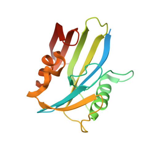
6EQ2, 6EQ3, 6EQ4, 6EQ5, 6EQ6, 6EQ7 - PubMed Abstract:
The DNA-repair enzyme MutT homolog 1 (MTH1) is a potential target for a broad range of tumors. Its substrate binding site features a non-catalytical pair of aspartic acids which resembles the catalytic dyad of aspartic proteases. We hypothesized that inhibitors of the latter might be re-targeted for MTH1 despite the two enzyme classes having different substrates and catalyze different reactions. We selected from the crystal structures of holo aspartic proteases a library of nearly 350 inhibitors for in silico screening. Three fragment hits were identified by docking and scoring according to a force field-based energy with continuum dielectric solvation. These fragments showed good ligand efficiency in a colorimetric assay (MW <300 Da and IC 50 <50μM). Molecular dynamics simulations were carried out for determining the most favorable interaction patterns. On the basis of the simulation results we evaluated in vitro seven commercially available compounds, two of which showed submicromolar potency for MTH1. To obtain definitive evidence of the predicted binding modes we solved the crystal structures of five of the 10 inhibitors predicted in silico. The final step of hit optimization was guided by protein crystallography and involved the synthesis of a single compound, the lead 11, which shows nanomolar affinity for MTH1 in two orthogonal binding assays, and selectivity higher than 2000-fold against its original target (BACE1). The high rate of fragment-hit identification and the fast optimization suggest that ligand retargeting by binding site analogy is an efficient strategy for drug design.
Organizational Affiliation:
Department of Biochemistry, University of Zurich, Winterthurerstrasse 190, CH-8057, Zurich, Switzerland.