6EN9

E. coli Hydrogenase-2 (hydrogen reduced form)


Experimental Data Snapshot

  • Method: X-RAY DIFFRACTION
  • Resolution: 1.50 Å
  • R-Value Free: 0.175 
  • R-Value Work: 0.152 
  • R-Value Observed: 0.153 

wwPDB Validation   3D Report Full Report


This is version 2.0 of the entry. See complete history


Literature

The structure of hydrogenase-2 fromEscherichia coli: implications for H2-driven proton pumping.

Beaton, S.E.Evans, R.M.Finney, A.J.Lamont, C.M.Armstrong, F.A.Sargent, F.Carr, S.B.

(2018) Biochem J 475: 1353-1370

  • DOI: https://doi.org/10.1042/BCJ20180053
  • Primary Citation of Related Structures:  
    6EHQ, 6EHS, 6EN9

  • PubMed Abstract: 

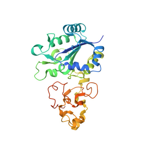
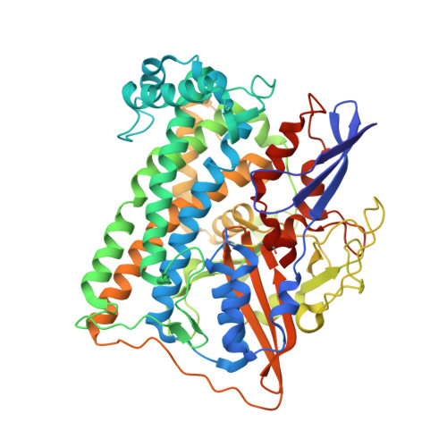
    Under anaerobic conditions, Escherichia coli is able to metabolize molecular hydrogen via the action of several [NiFe]-hydrogenase enzymes. Hydrogenase-2, which is typically present in cells at low levels during anaerobic respiration, is a periplasmic-facing membrane-bound complex that functions as a proton pump to convert energy from hydrogen (H 2 ) oxidation into a proton gradient; consequently, its structure is of great interest. Empirically, the complex consists of a tightly bound core catalytic module, comprising large (HybC) and small (HybO) subunits, which is attached to an Fe-S protein (HybA) and an integral membrane protein (HybB). To date, efforts to gain a more detailed picture have been thwarted by low native expression levels of Hydrogenase-2 and the labile interaction between HybOC and HybA/HybB subunits. In the present paper, we describe a new overexpression system that has facilitated the determination of high-resolution crystal structures of HybOC and, hence, a prediction of the quaternary structure of the HybOCAB complex.


  • Organizational Affiliation

    Department of Chemistry, Inorganic Chemistry Laboratory, University of Oxford, Oxford OX1 3QR, U.K.


Macromolecules
Find similar proteins by:  (by identity cutoff)  |  3D Structure
Entity ID: 1
MoleculeChains Sequence LengthOrganismDetailsImage
Hydrogenase-2 small chainA [auth S],
C [auth T]
298Escherichia coliMutation(s): 0 
Gene Names: hybOyghVb2997JW2965
EC: 1.12.99.6
UniProt
Find proteins for P69741 (Escherichia coli (strain K12))
Explore P69741 
Go to UniProtKB:  P69741
Entity Groups  
Sequence Clusters30% Identity50% Identity70% Identity90% Identity95% Identity100% Identity
UniProt GroupP69741
Sequence Annotations
Expand
  • Reference Sequence
Find similar proteins by:  (by identity cutoff)  |  3D Structure
Entity ID: 2
MoleculeChains Sequence LengthOrganismDetailsImage
Hydrogenase-2 large chainB [auth L],
D [auth M]
567Escherichia coliMutation(s): 0 
Gene Names: hybCb2994JW2962
EC: 1.12.99.6
UniProt
Find proteins for P0ACE0 (Escherichia coli (strain K12))
Explore P0ACE0 
Go to UniProtKB:  P0ACE0
Entity Groups  
Sequence Clusters30% Identity50% Identity70% Identity90% Identity95% Identity100% Identity
UniProt GroupP0ACE0
Sequence Annotations
Expand
  • Reference Sequence
Small Molecules
Ligands 6 Unique
IDChains Name / Formula / InChI Key2D Diagram3D Interactions
SF4
Query on SF4

Download Ideal Coordinates CCD File 
E [auth S],
G [auth S],
L [auth T],
N [auth T]
IRON/SULFUR CLUSTER
Fe4 S4
LJBDFODJNLIPKO-UHFFFAOYSA-N
F3S
Query on F3S

Download Ideal Coordinates CCD File 
F [auth S],
M [auth T]
FE3-S4 CLUSTER
Fe3 S4
FCXHZBQOKRZXKS-UHFFFAOYSA-N
FCO
Query on FCO

Download Ideal Coordinates CCD File 
I [auth L],
P [auth M]
CARBONMONOXIDE-(DICYANO) IRON
C3 Fe N2 O
VBQUCMTXYFMTTE-UHFFFAOYSA-N
SO4
Query on SO4

Download Ideal Coordinates CCD File 
H [auth S],
O [auth T]
SULFATE ION
O4 S
QAOWNCQODCNURD-UHFFFAOYSA-L
NI
Query on NI

Download Ideal Coordinates CCD File 
J [auth L],
Q [auth M]
NICKEL (II) ION
Ni
VEQPNABPJHWNSG-UHFFFAOYSA-N
MG
Query on MG

Download Ideal Coordinates CCD File 
K [auth L],
R [auth M]
MAGNESIUM ION
Mg
JLVVSXFLKOJNIY-UHFFFAOYSA-N
Experimental Data & Validation

Experimental Data

  • Method: X-RAY DIFFRACTION
  • Resolution: 1.50 Å
  • R-Value Free: 0.175 
  • R-Value Work: 0.152 
  • R-Value Observed: 0.153 
  • Space Group: P 21 21 21
Unit Cell:
Length ( Å )Angle ( ˚ )
a = 99.602α = 90
b = 100.54β = 90
c = 168.587γ = 90
Software Package:
Software NamePurpose
REFMACrefinement
DIALSdata reduction
Aimlessdata scaling
PHASERphasing

Structure Validation

View Full Validation Report


Entry History & Funding Information

Deposition Data


Funding OrganizationLocationGrant Number
Biotechnology and Biological Sciences Research CouncilUnited KingdomBB/N006321/1

Revision History  (Full details and data files)

  • Version 1.0: 2018-04-18
    Type: Initial release
  • Version 1.1: 2018-04-25
    Changes: Data collection, Database references
  • Version 2.0: 2024-01-17
    Changes: Atomic model, Data collection, Database references, Derived calculations, Refinement description, Structure summary