6EKO

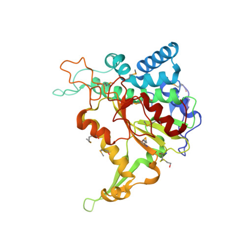
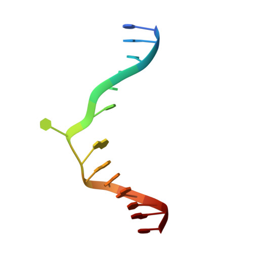
Crystal structure of Type IIP restriction endonuclease PfoI with cognate DNA


Experimental Data Snapshot

  • Method: X-RAY DIFFRACTION
  • Resolution: 2.28 Å
  • R-Value Free: 0.248 
  • R-Value Work: 0.205 
  • R-Value Observed: 0.209 

wwPDB Validation   3D Report Full Report


This is version 1.1 of the entry. See complete history


Literature

Unique mechanism of target recognition by PfoI restriction endonuclease of the CCGG-family.

Tamulaitiene, G.Manakova, E.Jovaisaite, V.Tamulaitis, G.Grazulis, S.Bochtler, M.Siksnys, V.

(2019) Nucleic Acids Res 47: 997-1010

  • DOI: https://doi.org/10.1093/nar/gky1137
  • Primary Citation of Related Structures:  
    6EK1, 6EKO

  • PubMed Abstract: 

    Restriction endonucleases (REs) of the CCGG-family recognize a set of 4-8 bp target sequences that share a common CCGG or CCNGG core and possess PD…D/ExK nuclease fold. REs that interact with 5 bp sequence 5'-CCNGG flip the central N nucleotides and 'compress' the bound DNA to stack the inner base pairs to mimic the CCGG sequence. PfoI belongs to the CCGG-family and cleaves the 7 bp sequence 5'-T|CCNGGA ("|" designates cleavage position). We present here crystal structures of PfoI in free and DNA-bound forms that show unique active site arrangement and mechanism of sequence recognition. Structures and mutagenesis indicate that PfoI features a permuted E…ExD…K active site that differs from the consensus motif characteristic to other family members. Although PfoI also flips the central N nucleotides of the target sequence it does not 'compress' the bound DNA. Instead, PfoI induces a drastic change in DNA backbone conformation that shortens the distance between scissile phosphates to match that in the unperturbed CCGG sequence. Our data demonstrate the diversity and versatility of structural mechanisms employed by restriction enzymes for recognition of related DNA sequences.


  • Organizational Affiliation

    Institute of Biotechnology, Vilnius University, Sauletekio al. 7, LT-10257 Vilnius, Lithuania.


Macromolecules

Find similar proteins by:  (by identity cutoff)  |  3D Structure
Entity ID: 1
MoleculeChains Sequence LengthOrganismDetailsImage
Restriction endonuclease PfoI
A, B
312Pseudomonas fluorescensMutation(s): 0 
EC: 3.1.21.4
UniProt
Find proteins for A0A452CST9 (Pseudomonas fluorescens)
Explore A0A452CST9 
Go to UniProtKB:  A0A452CST9
Entity Groups  
Sequence Clusters30% Identity50% Identity70% Identity90% Identity95% Identity100% Identity
UniProt GroupA0A452CST9
Sequence Annotations
Expand
  • Reference Sequence

Find similar nucleic acids by:  Sequence   |   3D Structure  

Entity ID: 2
MoleculeChains LengthOrganismImage
DNA (5'-D(*CP*GP*CP*TP*CP*CP*CP*GP*GP*AP*GP*CP*GP*T)-3')C [auth F],
D [auth E]
14Pseudomonas fluorescens
Sequence Annotations
Expand
  • Reference Sequence
Experimental Data & Validation

Experimental Data

  • Method: X-RAY DIFFRACTION
  • Resolution: 2.28 Å
  • R-Value Free: 0.248 
  • R-Value Work: 0.205 
  • R-Value Observed: 0.209 
  • Space Group: P 21 21 21
Unit Cell:
Length ( Å )Angle ( ˚ )
a = 56.279α = 90
b = 91.663β = 90
c = 152.742γ = 90
Software Package:
Software NamePurpose
XDSdata reduction
SCALAdata scaling
Auto-Rickshawphasing
Cootmodel building
PHENIXrefinement

Structure Validation

View Full Validation Report


Entry History & Funding Information

Deposition Data


Funding OrganizationLocationGrant Number
Research Council of LithuaniaLithuaniaMIP-41/2013
European UnionBioStruct-X (grant agreement 283570)

Revision History  (Full details and data files)

  • Version 1.0: 2018-10-10
    Type: Initial release
  • Version 1.1: 2019-04-24
    Changes: Data collection, Database references, Structure summary