6EJN

The KLC2 TPR domain bound to the JIP3 leucine zipper domain


Experimental Data Snapshot

  • Method: X-RAY DIFFRACTION
  • Resolution: 3.20 Å
  • R-Value Free: 0.210 
  • R-Value Work: 0.191 
  • R-Value Observed: 0.192 

wwPDB Validation   3D Report Full Report


This is version 1.3 of the entry. See complete history


Literature

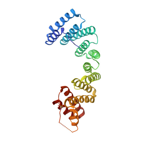
Insights into Kinesin-1 Activation from the Crystal Structure of KLC2 Bound to JIP3.

Cockburn, J.J.B.Hesketh, S.J.Mulhair, P.Thomsen, M.O'Connell, M.J.Way, M.

(2018) Structure 26: 1486-1498.e6

  • DOI: https://doi.org/10.1016/j.str.2018.07.011

  • PubMed Abstract: 

    Kinesin-1 transports numerous cellular cargoes along microtubules. The kinesin-1 light chain (KLC) mediates cargo binding and regulates kinesin-1 motility. To investigate the molecular basis for kinesin-1 recruitment and activation by cargoes, we solved the crystal structure of the KLC2 tetratricopeptide repeat (TPR) domain bound to the cargo JIP3. This, combined with biophysical and molecular evolutionary analyses, reveals a kinesin-1 cargo binding site, located on KLC TPR1, which is conserved in homologs from sponges to humans. In the complex, JIP3 crosslinks two KLC2 TPR domains via their TPR1s. We show that TPR1 forms a dimer interface that mimics JIP3 binding in all crystal structures of the unbound KLC TPR domain. We propose that cargo-induced dimerization of the KLC TPR domains via TPR1 is a general mechanism for activating kinesin-1. We relate this to activation by tryptophan-acidic cargoes, explaining how different cargoes activate kinesin-1 through related molecular mechanisms.


  • Organizational Affiliation

    Astbury Centre for Structural Molecular Biology, University of Leeds, Leeds LS2 9JT, UK. Electronic address: j.j.b.cockburn@leeds.ac.uk.


Macromolecules
Find similar proteins by:  (by identity cutoff)  |  3D Structure
Entity ID: 1
MoleculeChains Sequence LengthOrganismDetailsImage
Kinesin light chain 2
A, B
291Mus musculusMutation(s): 0 
Gene Names: Klc2mCG_8395
UniProt & NIH Common Fund Data Resources
Find proteins for O88448 (Mus musculus)
Explore O88448 
Go to UniProtKB:  O88448
IMPC:  MGI:107953
Entity Groups  
Sequence Clusters30% Identity50% Identity70% Identity90% Identity95% Identity100% Identity
UniProt GroupO88448
Sequence Annotations
Expand
  • Reference Sequence
Find similar proteins by:  (by identity cutoff)  |  3D Structure
Entity ID: 2
MoleculeChains Sequence LengthOrganismDetailsImage
C-Jun-amino-terminal kinase-interacting protein 3
C, D
75Mus musculusMutation(s): 0 
Gene Names: Mapk8ip3Jip3Jsap1Syd2
UniProt
Find proteins for Q9ESN9 (Mus musculus)
Explore Q9ESN9 
Go to UniProtKB:  Q9ESN9
Entity Groups  
Sequence Clusters30% Identity50% Identity70% Identity90% Identity95% Identity100% Identity
UniProt GroupQ9ESN9
Sequence Annotations
Expand
  • Reference Sequence
Experimental Data & Validation

Experimental Data

  • Method: X-RAY DIFFRACTION
  • Resolution: 3.20 Å
  • R-Value Free: 0.210 
  • R-Value Work: 0.191 
  • R-Value Observed: 0.192 
  • Space Group: P 65
Unit Cell:
Length ( Å )Angle ( ˚ )
a = 163.301α = 90
b = 163.301β = 90
c = 77.123γ = 120
Software Package:
Software NamePurpose
BUSTERrefinement
SCALAdata scaling
PHASERphasing
PDB_EXTRACTdata extraction

Structure Validation

View Full Validation Report


Entry History & Funding Information

Deposition Data


Funding OrganizationLocationGrant Number
Royal SocietyUnited KingdomRG150205

Revision History  (Full details and data files)

  • Version 1.0: 2018-09-05
    Type: Initial release
  • Version 1.1: 2018-09-19
    Changes: Data collection, Database references
  • Version 1.2: 2018-11-14
    Changes: Data collection, Database references
  • Version 1.3: 2024-01-17
    Changes: Data collection, Database references, Refinement description