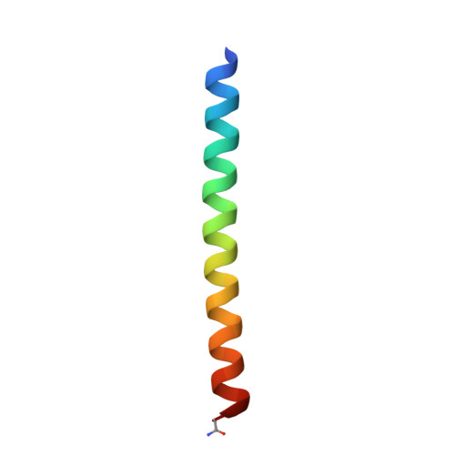
6EGN

Crystal Structure of a Three-stranded Coiled Coil Peptide Containing a Trigonal Planar Hg(II)S3 Site Modified by D-Leu in the Second Coordination Sphere


Experimental Data Snapshot

  • Method: X-RAY DIFFRACTION
  • Resolution: 1.84 Å
  • R-Value Free: 0.209 
  • R-Value Work: 0.197 
  • R-Value Observed: 0.198 

wwPDB Validation   3D Report Full Report


This is version 1.3 of the entry. See complete history


Literature

How Outer Coordination Sphere Modifications Can Impact Metal Structures in Proteins: A Crystallographic Evaluation.

Ruckthong, L.Stuckey, J.A.Pecoraro, V.L.

(2019) Chemistry 25: 6773-6787

  • DOI: https://doi.org/10.1002/chem.201806040
  • Primary Citation of Related Structures:  
    6EGL, 6EGM, 6EGN, 6EGO

  • PubMed Abstract: 

    A challenging objective of de novo metalloprotein design is to control of the outer coordination spheres of an active site to fine tune metal properties. The well-defined three stranded coiled coils, TRI and CoilSer peptides, are used to address this question. Substitution of Cys for Leu yields a thiophilic site within the core. Metals such as Hg II , Pb II , and As III result in trigonal planar or trigonal pyramidal geometries; however, spectroscopic studies have shown that Cd II forms three-, four- or five-coordinate Cd II S 3 (OH 2 ) x (in which x=0-2) when the outer coordination spheres are perturbed. Unfortunately, there has been little crystallographic examination of these proteins to explain the observations. Here, the high-resolution X-ray structures of apo- and mercurated proteins are compared to explain the modifications that lead to metal coordination number and geometry variation. It reveals that Ala substitution for Leu opens a cavity above the Cys site allowing for water excess, facilitating Cd II S 3 (OH 2 ). Replacement of Cys by Pen restricts thiol rotation, causing a shift in the metal-binding plane, which displaces water, forming Cd II S 3 . Residue d-Leu, above the Cys site, reorients the side chain towards the Cys layer, diminishing the space for water accommodation yielding Cd II S 3 , whereas d-Leu below opens more space, allowing for equal Cd II S 3 (OH 2 ) and Cd II S 3 (OH 2 ) 2 . These studies provide insights into how to control desired metal geometries in metalloproteins by using coded and non-coded amino acids.


  • Organizational Affiliation

    Department of Chemistry, University of Michigan, Ann Arbor, Michigan, 48109, USA.


Macromolecules
Find similar proteins by:  (by identity cutoff)  |  3D Structure
Entity ID: 1
MoleculeChains Sequence LengthOrganismDetailsImage
Hg(II)(GRAND CoilSerL16CL19(DLE))3-
A, B, C
38synthetic constructMutation(s): 0 
Entity Groups  
Sequence Clusters30% Identity50% Identity70% Identity90% Identity95% Identity100% Identity
Sequence Annotations
Expand
  • Reference Sequence
Small Molecules
Ligands 5 Unique
IDChains Name / Formula / InChI Key2D Diagram3D Interactions
15P
Query on 15P

Download Ideal Coordinates CCD File 
F [auth A],
G [auth A]
POLYETHYLENE GLYCOL (N=34)
C69 H140 O35
VUYXVWGKCKTUMF-UHFFFAOYSA-N
HG
Query on HG

Download Ideal Coordinates CCD File 
I [auth A]MERCURY (II) ION
Hg
BQPIGGFYSBELGY-UHFFFAOYSA-N
PRO
Query on PRO

Download Ideal Coordinates CCD File 
E [auth A],
K [auth B]
PROLINE
C5 H9 N O2
ONIBWKKTOPOVIA-BYPYZUCNSA-N
ZN
Query on ZN

Download Ideal Coordinates CCD File 
D [auth A],
J [auth B],
L [auth C]
ZINC ION
Zn
PTFCDOFLOPIGGS-UHFFFAOYSA-N
CL
Query on CL

Download Ideal Coordinates CCD File 
H [auth A]CHLORIDE ION
Cl
VEXZGXHMUGYJMC-UHFFFAOYSA-M
Experimental Data & Validation

Experimental Data

  • Method: X-RAY DIFFRACTION
  • Resolution: 1.84 Å
  • R-Value Free: 0.209 
  • R-Value Work: 0.197 
  • R-Value Observed: 0.198 
  • Space Group: P 21 21 21
Unit Cell:
Length ( Å )Angle ( ˚ )
a = 32.636α = 90
b = 80.508β = 90
c = 88.73γ = 90
Software Package:
Software NamePurpose
HKL-2000data scaling
BUSTER-TNTrefinement
PDB_EXTRACTdata extraction
HKL-2000data reduction
AutoSolphasing
HKL-2000data collection

Structure Validation

View Full Validation Report


Entry History & Funding Information

Deposition Data


Funding OrganizationLocationGrant Number
National Institutes of Health/National Human Genome Research Institute (NIH/NHGRI)United StatesR01ES012236

Revision History  (Full details and data files)

  • Version 1.0: 2019-04-03
    Type: Initial release
  • Version 1.1: 2019-05-08
    Changes: Data collection, Database references
  • Version 1.2: 2019-05-22
    Changes: Data collection, Database references
  • Version 1.3: 2019-12-18
    Changes: Author supporting evidence