6E8E

Crystal structure of the Escherichia coli sliding clamp-MutL complex.


Experimental Data Snapshot

  • Method: X-RAY DIFFRACTION
  • Resolution: 2.25 Å
  • R-Value Free: 0.225 
  • R-Value Work: 0.190 
  • R-Value Observed: 0.191 

wwPDB Validation   3D Report Full Report


This is version 1.4 of the entry. See complete history


Literature

Binding of the regulatory domain of MutL to the sliding beta-clamp is species specific.

Almawi, A.W.Scotland, M.K.Randall, J.R.Liu, L.Martin, H.K.Sacre, L.Shen, Y.Pillon, M.C.Simmons, L.A.Sutton, M.D.Guarne, A.

(2019) Nucleic Acids Res 47: 4831-4842

  • DOI: https://doi.org/10.1093/nar/gkz115
  • Primary Citation of Related Structures:  
    6E8D, 6E8E

  • PubMed Abstract: 

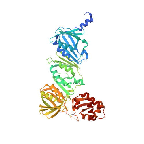
    The β-clamp is a protein hub central to DNA replication and fork management. Proteins interacting with the β-clamp harbor a conserved clamp-binding motif that is often found in extended regions. Therefore, clamp interactions have -almost exclusively- been studied using short peptides recapitulating the binding motif. This approach has revealed the molecular determinants that mediate the binding but cannot describe how proteins with clamp-binding motifs embedded in structured domains are recognized. The mismatch repair protein MutL has an internal clamp-binding motif, but its interaction with the β-clamp has different roles depending on the organism. In Bacillus subtilis, the interaction stimulates the endonuclease activity of MutL and it is critical for DNA mismatch repair. Conversely, disrupting the interaction between Escherichia coli MutL and the β-clamp only causes a mild mutator phenotype. Here, we determined the structures of the regulatory domains of E. coli and B. subtilis MutL bound to their respective β-clamps. The structures reveal different binding modes consistent with the binding to the β-clamp being a two-step process. Functional characterization indicates that, within the regulatory domain, only the clamp binding motif is required for the interaction between the two proteins. However, additional motifs beyond the regulatory domain may stabilize the interaction. We propose a model for the activation of the endonuclease activity of MutL in organisms lacking methyl-directed mismatch repair.


  • Organizational Affiliation

    Department of Biochemistry and Biomedical Sciences, McMaster University, Hamilton, ON, Canada.


Macromolecules
Find similar proteins by:  (by identity cutoff)  |  3D Structure
Entity ID: 1
MoleculeChains Sequence LengthOrganismDetailsImage
Beta sliding clamp,DNA mismatch repair protein MutL517Escherichia coli K-12Escherichia coli O139:H28 str. E24377A
This entity is chimeric
Mutation(s): 0 
Gene Names: dnaNb3701JW3678mutLEcE24377A_4728
UniProt
Find proteins for P0A988 (Escherichia coli (strain K12))
Explore P0A988 
Go to UniProtKB:  P0A988
Find proteins for P23367 (Escherichia coli (strain K12))
Explore P23367 
Go to UniProtKB:  P23367
Entity Groups  
Sequence Clusters30% Identity50% Identity70% Identity90% Identity95% Identity100% Identity
UniProt GroupsP23367P0A988
Sequence Annotations
Expand
  • Reference Sequence
Find similar proteins by:  (by identity cutoff)  |  3D Structure
Entity ID: 2
MoleculeChains Sequence LengthOrganismDetailsImage
Beta sliding clamp,DNA mismatch repair protein MutL517Escherichia coli K-12Escherichia coli O139:H28 str. E24377A
This entity is chimeric
Mutation(s): 0 
Gene Names: dnaNb3701JW3678mutLEcE24377A_4728
UniProt
Find proteins for P0A988 (Escherichia coli (strain K12))
Explore P0A988 
Go to UniProtKB:  P0A988
Find proteins for P23367 (Escherichia coli (strain K12))
Explore P23367 
Go to UniProtKB:  P23367
Entity Groups  
Sequence Clusters30% Identity50% Identity70% Identity90% Identity95% Identity100% Identity
UniProt GroupsP23367P0A988
Sequence Annotations
Expand
  • Reference Sequence
Small Molecules
Ligands 2 Unique
IDChains Name / Formula / InChI Key2D Diagram3D Interactions
SO4
Query on SO4

Download Ideal Coordinates CCD File 
G [auth A],
H [auth A],
I [auth A],
L [auth B],
M [auth B]
SULFATE ION
O4 S
QAOWNCQODCNURD-UHFFFAOYSA-L
GOL
Query on GOL

Download Ideal Coordinates CCD File 
C [auth A]
D [auth A]
E [auth A]
F [auth A]
J [auth B]
C [auth A],
D [auth A],
E [auth A],
F [auth A],
J [auth B],
K [auth B]
GLYCEROL
C3 H8 O3
PEDCQBHIVMGVHV-UHFFFAOYSA-N
Modified Residues  1 Unique
IDChains TypeFormula2D DiagramParent
CSO
Query on CSO
A
L-PEPTIDE LINKINGC3 H7 N O3 SCYS
Experimental Data & Validation

Experimental Data

  • Method: X-RAY DIFFRACTION
  • Resolution: 2.25 Å
  • R-Value Free: 0.225 
  • R-Value Work: 0.190 
  • R-Value Observed: 0.191 
  • Space Group: P 21 21 21
Unit Cell:
Length ( Å )Angle ( ˚ )
a = 78.39α = 90
b = 103.21β = 90
c = 141.64γ = 90
Software Package:
Software NamePurpose
PHENIXrefinement
XDSdata reduction
XDSdata scaling
PHASERphasing

Structure Validation

View Full Validation Report


Entry History 

Deposition Data

Revision History  (Full details and data files)

  • Version 1.0: 2019-05-01
    Type: Initial release
  • Version 1.1: 2019-05-22
    Changes: Data collection, Database references
  • Version 1.2: 2020-10-07
    Changes: Derived calculations
  • Version 1.3: 2023-10-11
    Changes: Data collection, Database references, Refinement description
  • Version 1.4: 2023-11-15
    Changes: Data collection