6DU7

Glutathione reductase from Streptococcus pneumoniae


Experimental Data Snapshot

  • Method: X-RAY DIFFRACTION
  • Resolution: 2.56 Å
  • R-Value Free: 0.233 
  • R-Value Work: 0.180 
  • R-Value Observed: 0.183 

wwPDB Validation   3D Report Full Report


Ligand Structure Quality Assessment 


This is version 1.3 of the entry. See complete history


Literature

The structure and activity of the glutathione reductase from Streptococcus pneumoniae.

Sikanyika, M.Aragao, D.McDevitt, C.A.Maher, M.J.

(2019) Acta Crystallogr F Struct Biol Commun 75: 54-61

  • DOI: https://doi.org/10.1107/S2053230X18016527
  • Primary Citation of Related Structures:  
    6DU7

  • PubMed Abstract: 

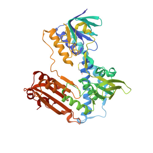
    The glutathione reductase (GR) from Streptococcus pneumoniae is a flavoenzyme that catalyzes the reduction of oxidized glutathione (GSSG) to its reduced form (GSH) in the cytoplasm of this bacterium. The maintenance of an intracellular pool of GSH is critical for the detoxification of reactive oxygen and nitrogen species and for intracellular metal tolerance to ions such as zinc. Here, S. pneumoniae GR (SpGR) was overexpressed and purified and its crystal structure determined at 2.56 Å resolution. SpGR shows overall structural similarity to other characterized GRs, with a dimeric structure that includes an antiparallel β-sheet at the dimer interface. This observation, in conjunction with comparisons with the interface structures of other GR enzymes, allows the classification of these enzymes into three classes. Analyses of the kinetic properties of SpGR revealed a significantly higher value for K m(GSSG) (231.2 ± 24.7 µM) in comparison to other characterized GR enzymes.


  • Organizational Affiliation

    Department of Biochemistry and Genetics, La Trobe Institute for Molecular Science, La Trobe University, Melbourne 3086, Australia.


Macromolecules
Find similar proteins by:  (by identity cutoff)  |  3D Structure
Entity ID: 1
MoleculeChains Sequence LengthOrganismDetailsImage
Glutathione reductase
A, B, C, D, E
A, B, C, D, E, F, G, H
448Streptococcus pneumoniaeMutation(s): 0 
Gene Names: gorA5N45_09080ERS020408_00588ERS022363_01056
EC: 1.8.1.7
UniProt
Find proteins for A0A0U0ARX4 (Streptococcus pneumoniae)
Explore A0A0U0ARX4 
Go to UniProtKB:  A0A0U0ARX4
Entity Groups  
Sequence Clusters30% Identity50% Identity70% Identity90% Identity95% Identity100% Identity
UniProt GroupA0A0U0ARX4
Sequence Annotations
Expand
  • Reference Sequence
Small Molecules
Ligands 2 Unique
IDChains Name / Formula / InChI Key2D Diagram3D Interactions
FAD
Query on FAD

Download Ideal Coordinates CCD File 
I [auth A]
J [auth B]
L [auth C]
M [auth D]
N [auth E]
I [auth A],
J [auth B],
L [auth C],
M [auth D],
N [auth E],
P [auth F],
Q [auth G],
R [auth H]
FLAVIN-ADENINE DINUCLEOTIDE
C27 H33 N9 O15 P2
VWWQXMAJTJZDQX-UYBVJOGSSA-N
SCN
Query on SCN

Download Ideal Coordinates CCD File 
K [auth B],
O [auth E]
THIOCYANATE ION
C N S
ZMZDMBWJUHKJPS-UHFFFAOYSA-M
Experimental Data & Validation

Experimental Data

  • Method: X-RAY DIFFRACTION
  • Resolution: 2.56 Å
  • R-Value Free: 0.233 
  • R-Value Work: 0.180 
  • R-Value Observed: 0.183 
  • Space Group: P 1 21 1
Unit Cell:
Length ( Å )Angle ( ˚ )
a = 96.748α = 90
b = 172.671β = 91.05
c = 135.434γ = 90
Software Package:
Software NamePurpose
REFMACrefinement
PDB_EXTRACTdata extraction
XDSdata reduction
Aimlessdata scaling
PHASERphasing

Structure Validation

View Full Validation Report


Ligand Structure Quality Assessment 


Entry History & Funding Information

Deposition Data


Funding OrganizationLocationGrant Number
National Health and Medical Research Council (NHMRC, Australia)AustraliaGNT1080784

Revision History  (Full details and data files)

  • Version 1.0: 2019-01-16
    Type: Initial release
  • Version 1.1: 2019-04-17
    Changes: Author supporting evidence, Data collection
  • Version 1.2: 2020-01-08
    Changes: Author supporting evidence
  • Version 1.3: 2023-10-11
    Changes: Data collection, Database references, Refinement description