6DKC

Yeast Ddi2 Cyanamide Hydratase, T157V mutant, apo structure


Experimental Data Snapshot

  • Method: X-RAY DIFFRACTION
  • Resolution: 2.90 Å
  • R-Value Free: 0.215 
  • R-Value Work: 0.193 
  • R-Value Observed: 0.193 

wwPDB Validation   3D Report Full Report


This is version 1.3 of the entry. See complete history


Literature

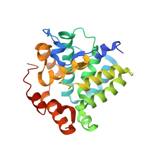
Structure of Ddi2, a highly inducible detoxifying metalloenzyme fromSaccharomyces cerevisiae.

Li, J.Jia, Y.Lin, A.Hanna, M.Chelico, L.Xiao, W.Moore, S.A.

(2019) J Biol Chem 294: 10674-10685

  • DOI: https://doi.org/10.1074/jbc.RA118.006394
  • Primary Citation of Related Structures:  
    6DK9, 6DKA, 6DKC, 6DKD

  • PubMed Abstract: 

    Cyanamide (H 2 N-CN) is used to break bud dormancy in woody plants and to deter alcohol use in humans. The biological effects of cyanamide in both these cases require the enzyme catalase. We previously demonstrated that Saccharomyces cerevisiae exposed to cyanamide resulted in strong induction of DDI2 gene expression. Ddi2 enzymatically hydrates cyanamide to urea and belongs to the family of HD-domain metalloenzymes (named after conserved active-site metal-binding His and Asp residues). Here, we report the X-ray structure of yeast Ddi2 to 2.6 Å resolution, revealing that Ddi2 is a dimeric zinc metalloenzyme. We also confirm that Ddi2 shares structural similarity with other known HD-domain proteins. HD residues His-55, His-88, and Asp-89 coordinate the active-site zinc, and the fourth zinc ligand is a water/hydroxide molecule. Other HD domain enzymes have a second aspartate metal ligand, but in Ddi2 this residue (Thr-157) does not interact with the zinc ion. Several Ddi2 active-site point mutations exhibited reduced catalytic activity. We kinetically and structurally characterized H137N and T157V mutants of Ddi2. A cyanamide soak of the Ddi2-T157V enzyme revealed cyanamide bound directly to the Zn 2+ ion, having displaced the zinc-bound water molecule. The mode of cyanamide binding to Ddi2 resembles cyanamide binding to the active-site zinc of carbonic anhydrase, a known cyanamide hydratase. Finally, we observed that the sensitivity of ddi2 Δ ddi3 Δ to cyanamide was not rescued by plasmids harboring ddi2-H137N or ddi2-TI57V variants, demonstrating that yeast cells require a functioning cyanamide hydratase to overcome cyanamide-induced growth defects.


  • Organizational Affiliation

    From the Department of Biochemistry, Microbiology and Immunology, University of Saskatchewan, Saskatoon, Saskatchewan S7N 5E5, Canada and.


Macromolecules
Find similar proteins by:  (by identity cutoff)  |  3D Structure
Entity ID: 1
MoleculeChains Sequence LengthOrganismDetailsImage
DNA damage-inducible protein
A, B, C, D, E
A, B, C, D, E, F, G, H, I
234Saccharomyces cerevisiae YJM789Mutation(s): 1 
Gene Names: SCY_1694
EC: 4.2.1.69
UniProt
Find proteins for A7A1Y4 (Saccharomyces cerevisiae (strain YJM789))
Explore A7A1Y4 
Go to UniProtKB:  A7A1Y4
Entity Groups  
Sequence Clusters30% Identity50% Identity70% Identity90% Identity95% Identity100% Identity
UniProt GroupA7A1Y4
Sequence Annotations
Expand
  • Reference Sequence
Small Molecules
Ligands 2 Unique
IDChains Name / Formula / InChI Key2D Diagram3D Interactions
SO4
Query on SO4

Download Ideal Coordinates CCD File 
AA [auth D]
CA [auth D]
DA [auth D]
EA [auth D]
FA [auth D]
AA [auth D],
CA [auth D],
DA [auth D],
EA [auth D],
FA [auth D],
GA [auth D],
HA [auth D],
JA [auth E],
K [auth A],
KA [auth E],
L [auth A],
LA [auth E],
M [auth A],
MA [auth E],
N [auth B],
NA [auth E],
OA [auth E],
P [auth B],
PA [auth F],
Q [auth B],
R [auth B],
RA [auth F],
S [auth B],
SA [auth F],
U [auth C],
UA [auth G],
V [auth C],
VA [auth G],
W [auth C],
X [auth C],
Y [auth C],
Z [auth C]
SULFATE ION
O4 S
QAOWNCQODCNURD-UHFFFAOYSA-L
ZN (Subject of Investigation/LOI)
Query on ZN

Download Ideal Coordinates CCD File 
BA [auth D]
IA [auth E]
J [auth A]
O [auth B]
QA [auth F]
BA [auth D],
IA [auth E],
J [auth A],
O [auth B],
QA [auth F],
T [auth C],
TA [auth G],
WA [auth H],
XA [auth I]
ZINC ION
Zn
PTFCDOFLOPIGGS-UHFFFAOYSA-N
Experimental Data & Validation

Experimental Data

  • Method: X-RAY DIFFRACTION
  • Resolution: 2.90 Å
  • R-Value Free: 0.215 
  • R-Value Work: 0.193 
  • R-Value Observed: 0.193 
  • Space Group: P 3 2 1
Unit Cell:
Length ( Å )Angle ( ˚ )
a = 263.613α = 90
b = 263.613β = 90
c = 119.026γ = 120
Software Package:
Software NamePurpose
PHENIXrefinement
HKL-2000data reduction
HKL-2000data scaling
PHENIXphasing

Structure Validation

View Full Validation Report


Entry History & Funding Information

Deposition Data


Funding OrganizationLocationGrant Number
Natural Sciences and Engineering Research Council (NSERC, Canada)CanadaRGPIN-2014-04580
Natural Sciences and Engineering Research Council (NSERC, Canada)CanadaRGPIN-2011-262138
Canadian Institutes of Health Research (CIHR)CanadaMOP-126155

Revision History  (Full details and data files)

  • Version 1.0: 2019-05-08
    Type: Initial release
  • Version 1.1: 2019-11-27
    Changes: Database references
  • Version 1.2: 2020-01-08
    Changes: Author supporting evidence
  • Version 1.3: 2023-10-11
    Changes: Data collection, Database references, Derived calculations, Refinement description