6DEJ

The structure of HcRed7, a brighter and red-shifted HcRed variant


Experimental Data Snapshot

  • Method: X-RAY DIFFRACTION
  • Resolution: 1.63 Å
  • R-Value Free: 0.214 
  • R-Value Work: 0.178 

wwPDB Validation   3D Report Full Report


This is version 2.0 of the entry. See complete history


Literature

Monomerization of far-red fluorescent proteins.

Wannier, T.M.Gillespie, S.K.Hutchins, N.McIsaac, R.S.Wu, S.Y.Shen, Y.Campbell, R.E.Brown, K.S.Mayo, S.L.

(2018) Proc Natl Acad Sci U S A 115: E11294-E11301

  • DOI: https://doi.org/10.1073/pnas.1807449115
  • Primary Citation of Related Structures:  
    6DEJ

  • PubMed Abstract: 

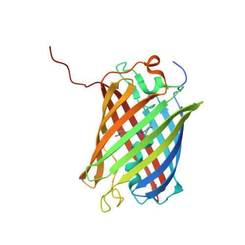
    Anthozoa -class red fluorescent proteins (RFPs) are frequently used as biological markers, with far-red (λ em ∼ 600-700 nm) emitting variants sought for whole-animal imaging because biological tissues are more permeable to light in this range. A barrier to the use of naturally occurring RFP variants as molecular markers is that all are tetrameric, which is not ideal for cell biological applications. Efforts to engineer monomeric RFPs have typically produced dimmer and blue-shifted variants because the chromophore is sensitive to small structural perturbations. In fact, despite much effort, only four native RFPs have been successfully monomerized, leaving the majority of RFP biodiversity untapped in biomarker development. Here we report the generation of monomeric variants of HcRed and mCardinal, both far-red dimers, and describe a comprehensive methodology for the monomerization of red-shifted oligomeric RFPs. Among the resultant variants is mKelly1 (emission maximum, λ em = 656 nm), which, along with the recently reported mGarnet2 [Matela G, et al. (2017) Chem Commun (Camb) 53:979-982], forms a class of bright, monomeric, far-red FPs.


  • Organizational Affiliation

    Division of Biology and Biological Engineering, California Institute of Technology, Pasadena, CA 91125; timothy_wannier@hms.harvard.edu steve@mayo.caltech.edu.


Macromolecules
Find similar proteins by:  (by identity cutoff)  |  3D Structure
Entity ID: 1
MoleculeChains Sequence LengthOrganismDetailsImage
GFP-like non-fluorescent chromoprotein
A, B, C, D
235Radianthus crispaMutation(s): 9 
UniProt
Find proteins for Q95W85 (Heteractis crispa)
Explore Q95W85 
Go to UniProtKB:  Q95W85
Entity Groups  
Sequence Clusters30% Identity50% Identity70% Identity90% Identity95% Identity100% Identity
UniProt GroupQ95W85
Sequence Annotations
Expand
  • Reference Sequence
Small Molecules
Experimental Data & Validation

Experimental Data

  • Method: X-RAY DIFFRACTION
  • Resolution: 1.63 Å
  • R-Value Free: 0.214 
  • R-Value Work: 0.178 
  • Space Group: P 1 21 1
Unit Cell:
Length ( Å )Angle ( ˚ )
a = 54.342α = 90
b = 122.057β = 108.75
c = 75.254γ = 90
Software Package:
Software NamePurpose
PHENIXrefinement
MOSFLMdata reduction
SCALAdata scaling
PHENIXphasing
Web-Icedata collection
Cootmodel building

Structure Validation

View Full Validation Report


Entry History & Funding Information

Deposition Data


Funding OrganizationLocationGrant Number
National Institutes of Health/National Institute of Biomedical Imaging and Bioengineering (NIH/NIBIB)United StatesR21EB018579

Revision History  (Full details and data files)

  • Version 1.0: 2018-05-23
    Type: Initial release
  • Version 1.1: 2018-12-05
    Changes: Data collection, Database references
  • Version 1.2: 2018-12-12
    Changes: Data collection, Database references
  • Version 1.3: 2019-12-11
    Changes: Author supporting evidence
  • Version 2.0: 2023-11-15
    Changes: Atomic model, Data collection, Database references, Derived calculations