


Crystal structure of Kynurenine Aminotransferase-II in apo-form
Jayawickrama, G.S., Sun, G., Nematollahi, A., Church, W.B.To be published.
Experimental Data Snapshot
wwPDB Validation 3D Report Full Report

Entity ID: 1 | |||||
|---|---|---|---|---|---|
| Molecule | Chains | Sequence Length | Organism | Details | Image |
| Kynurenine/alpha-aminoadipate aminotransferase, mitochondrial | 431 | Homo sapiens | Mutation(s): 0 Gene Names: AADAT, KAT2 EC: 2.6.1.39 (PDB Primary Data), 2.6.1.7 (PDB Primary Data) | 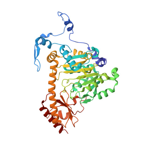 | |
UniProt & NIH Common Fund Data Resources | |||||
Find proteins for Q8N5Z0 (Homo sapiens) Explore Q8N5Z0 Go to UniProtKB: Q8N5Z0 | |||||
PHAROS: Q8N5Z0 GTEx: ENSG00000109576 | |||||
Entity Groups | |||||
| Sequence Clusters | 30% Identity50% Identity70% Identity90% Identity95% Identity100% Identity | ||||
| UniProt Group | Q8N5Z0 | ||||
Sequence AnnotationsExpand | |||||
| |||||
| Length ( Å ) | Angle ( ˚ ) |
|---|---|
| a = 102.765 | α = 90 |
| b = 102.765 | β = 90 |
| c = 86.498 | γ = 90 |
| Software Name | Purpose |
|---|---|
| phenix.refine | refinement |
| PHENIX | refinement |
| XDS | data reduction |
| Aimless | data scaling |
| PHENIX | phasing |
RCSB PDB (citation) is hosted by
RCSB PDB is a member of the

