6CIH

Crystal structure of a group II intron lariat in the post-catalytic state


Experimental Data Snapshot

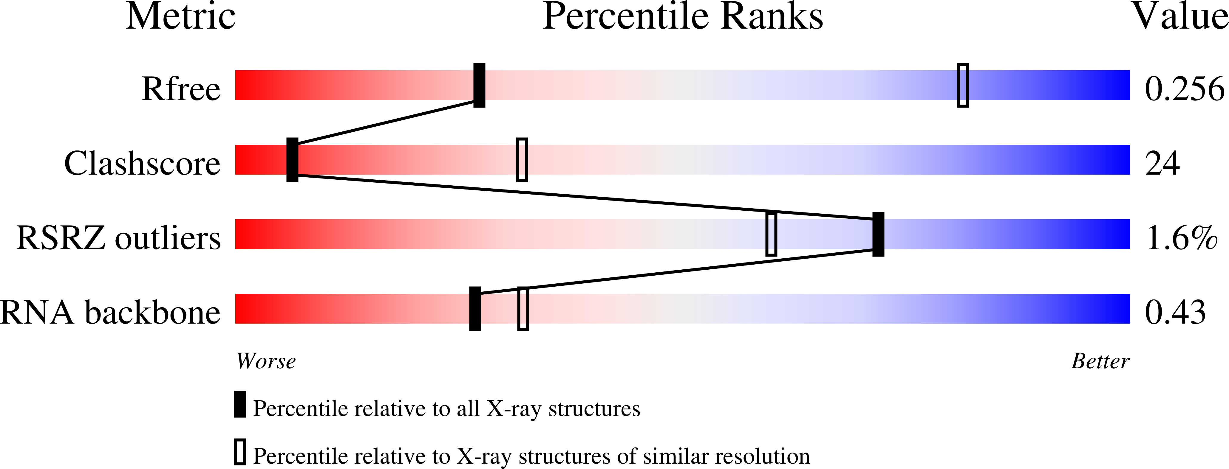
  • Method: X-RAY DIFFRACTION
  • Resolution: 3.68 Å
  • R-Value Free: 0.255 
  • R-Value Work: 0.208 
  • R-Value Observed: 0.211 

wwPDB Validation   3D Report Full Report


This is version 1.2 of the entry. See complete history

Re-refinement Note

This entry reflects an alternative modeling of the original data in: 4R0D


Literature

Structural basis for the second step of group II intron splicing.

Chan, R.T.Peters, J.K.Robart, A.R.Wiryaman, T.Rajashankar, K.R.Toor, N.

(2018) Nat Commun 9: 4676-4676

  • DOI: https://doi.org/10.1038/s41467-018-06678-0
  • Primary Citation of Related Structures:  
    6CHR, 6CIH

  • PubMed Abstract: 

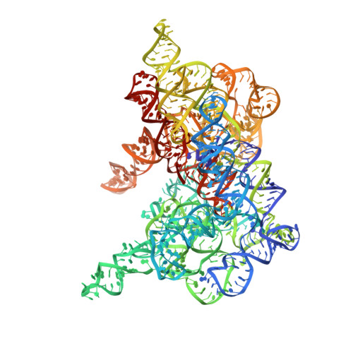
    The group II intron and the spliceosome share a common active site architecture and are thought to be evolutionarily related. Here we report the 3.7 Å crystal structure of a eukaryotic group II intron in the lariat-3' exon form, immediately preceding the second step of splicing, analogous to the spliceosomal P complex. This structure reveals the location of the intact 3' splice site within the catalytic core of the group II intron. The 3'-OH of the 5' exon is positioned in close proximity to the 3' splice site for nucleophilic attack and exon ligation. The active site undergoes conformational rearrangements with the catalytic triplex having different configurations before and after the second step of splicing. We describe a complete model for the second step of group II intron splicing that incorporates a dynamic catalytic triplex being responsible for creating the binding pocket for 3' splice site capture.


  • Organizational Affiliation

    Department of Chemistry and Biochemistry, University of California, San Diego, La Jolla, CA, 92093, USA.


Macromolecules
Find similar nucleic acids by:  (by identity cutoff)  |  3D Structure
Entity ID: 1
MoleculeChains LengthOrganismImage
RNA (696-MER)621Pylaiella littoralis
Sequence Annotations
Expand
  • Reference Sequence

Find similar nucleic acids by:  Sequence   |   3D Structure  

Entity ID: 2
MoleculeChains LengthOrganismImage
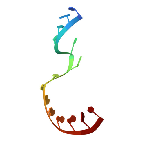
RNA (5'-R(P*UP*GP*UP*UP*UP*AP*UP*UP*AP*AP*AP*AP*AP*C*-3')14Pylaiella littoralis
Sequence Annotations
Expand
  • Reference Sequence
Small Molecules
Ligands 3 Unique
IDChains Name / Formula / InChI Key2D Diagram3D Interactions
IRI
Query on IRI

Download Ideal Coordinates CCD File 
AA [auth A]
BA [auth A]
C [auth A]
CA [auth A]
D [auth A]
AA [auth A],
BA [auth A],
C [auth A],
CA [auth A],
D [auth A],
DA [auth A],
E [auth A],
EA [auth A],
F [auth A],
FA [auth A],
G [auth A],
GA [auth A],
H [auth A],
HA [auth A],
I [auth A],
IA [auth A],
J [auth A],
JA [auth A],
K [auth A],
KA [auth A],
L [auth A],
LA [auth A],
M [auth A],
N [auth A],
O [auth A],
P [auth A],
Q [auth A],
R [auth A],
S [auth A],
T [auth A],
U [auth A],
UD [auth A],
V [auth A],
VD [auth A],
W [auth A],
WD [auth A],
X [auth A],
XD [auth A],
Y [auth A],
YD [auth A],
Z [auth A]
IRIDIUM HEXAMMINE ION
H18 Ir N6
CGMAOQLDNKCXGK-UHFFFAOYSA-N
MG
Query on MG

Download Ideal Coordinates CCD File 
AB [auth A]
AD [auth A]
AE [auth A]
BB [auth A]
BD [auth A]
AB [auth A],
AD [auth A],
AE [auth A],
BB [auth A],
BD [auth A],
BE [auth A],
CB [auth A],
CD [auth A],
CE [auth A],
DB [auth A],
DD [auth A],
DE [auth A],
EB [auth A],
EC [auth A],
ED [auth A],
EE [auth A],
FB [auth A],
FC [auth A],
FD [auth A],
FE [auth A],
GB [auth A],
GC [auth A],
GD [auth A],
GE [auth B],
HB [auth A],
HC [auth A],
HD [auth A],
HE [auth B],
IB [auth A],
IC [auth A],
ID [auth A],
JB [auth A],
JC [auth A],
JD [auth A],
KB [auth A],
KC [auth A],
KD [auth A],
LB [auth A],
LC [auth A],
LD [auth A],
MA [auth A],
MB [auth A],
MC [auth A],
MD [auth A],
NA [auth A],
NB [auth A],
NC [auth A],
ND [auth A],
OA [auth A],
OB [auth A],
OC [auth A],
OD [auth A],
PA [auth A],
PB [auth A],
PC [auth A],
PD [auth A],
QA [auth A],
QB [auth A],
QC [auth A],
QD [auth A],
RA [auth A],
RB [auth A],
RC [auth A],
RD [auth A],
SA [auth A],
SB [auth A],
SC [auth A],
SD [auth A],
TA [auth A],
TB [auth A],
TC [auth A],
TD [auth A],
UA [auth A],
UB [auth A],
UC [auth A],
VA [auth A],
VC [auth A],
WA [auth A],
WC [auth A],
XA [auth A],
XC [auth A],
YA [auth A],
YC [auth A],
ZA [auth A],
ZC [auth A],
ZD [auth A]
MAGNESIUM ION
Mg
JLVVSXFLKOJNIY-UHFFFAOYSA-N
NA
Query on NA

Download Ideal Coordinates CCD File 
AC [auth A]
BC [auth A]
CC [auth A]
DC [auth A]
VB [auth A]
AC [auth A],
BC [auth A],
CC [auth A],
DC [auth A],
VB [auth A],
WB [auth A],
XB [auth A],
YB [auth A],
ZB [auth A]
SODIUM ION
Na
FKNQFGJONOIPTF-UHFFFAOYSA-N
Experimental Data & Validation

Experimental Data

  • Method: X-RAY DIFFRACTION
  • Resolution: 3.68 Å
  • R-Value Free: 0.255 
  • R-Value Work: 0.208 
  • R-Value Observed: 0.211 
  • Space Group: C 2 2 21
Unit Cell:
Length ( Å )Angle ( ˚ )
a = 163.639α = 90
b = 255.361β = 90
c = 136.786γ = 90
Software Package:
Software NamePurpose
PHENIXrefinement
HKL-2000data reduction
HKL-2000data scaling
SHELXDEphasing

Structure Validation

View Full Validation Report


Entry History & Funding Information

Deposition Data


Funding OrganizationLocationGrant Number
National Institutes of Health/National Institute of General Medical Sciences (NIH/NIGMS)United States5R01GM102216
National Institutes of Health/National Institute of General Medical Sciences (NIH/NIGMS)United States1R01GM123275
National Institutes of Health/National Institute of General Medical Sciences (NIH/NIGMS)United States5P41GM103403
National Institutes of Health/National Institute of General Medical Sciences (NIH/NIGMS)United States5T32GM007240
National Institutes of Health/National Institute of General Medical Sciences (NIH/NIGMS)United States5T32GM008326

Revision History  (Full details and data files)

  • Version 1.0: 2018-11-21
    Type: Initial release
  • Version 1.1: 2020-01-01
    Changes: Author supporting evidence
  • Version 1.2: 2024-03-13
    Changes: Data collection, Database references, Derived calculations