6C9L

MEF2B Apo Protein Structure

  • Classification: TRANSCRIPTION
  • Organism(s): Homo sapiens
  • Expression System: Escherichia coli
  • Mutation(s): No 

  • Deposited: 2018-01-26 Released: 2018-02-07 
  • Deposition Author(s): Lei, X., Chen, L.
  • Funding Organization(s): National Institutes of Health/National Human Genome Research Institute (NIH/NHGRI)

Experimental Data Snapshot

  • Method: X-RAY DIFFRACTION
  • Resolution: 2.30 Å
  • R-Value Free: 0.235 
  • R-Value Work: 0.215 
  • R-Value Observed: 0.216 

wwPDB Validation   3D Report Full Report


This is version 1.4 of the entry. See complete history


Literature

Crystal Structure of Apo MEF2B Reveals New Insights in DNA Binding and Cofactor Interaction.

Lei, X.Shi, H.Kou, Y.Rajashekar, N.Wu, F.Sen, C.Xu, J.Chen, L.

(2018) Biochemistry 57: 4047-4051

  • DOI: https://doi.org/10.1021/acs.biochem.8b00439
  • Primary Citation of Related Structures:  
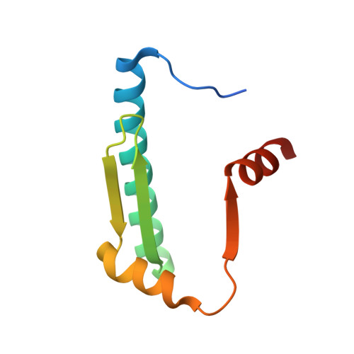
    6C9L

  • PubMed Abstract: 

    The myocyte enhancer factor 2 (MEF2) family of transcription factors plays important roles in developmental processes and adaptive responses. Although MEF2 proteins are known to bind DNA in the nucleus to regulate specific gene expression, there are reports that show that MEF2 also functions in the cytoplasm. Previous structural studies of MEF2 focused exclusively on DNA-bound MEF2 with and without various corepressors or coactivators. While these studies have established a comprehensive structural model of DNA recognition and cofactor recruitment by MEF2, the structure of MEF2 not bound to DNA, which include cytoplasmic MEF2 and free MEF2 in the nucleus, is unknown. Here we determined the structure of the MADS-box/MEF2 domain of MEF2B without DNA nor cofactor. The Apo structure of MEF2B reveals a largely preformed DNA binding interface that may be important for recognizing the shape of DNA from the minor groove side. In addition, our structure also reveals that the C-terminal helix of the MEF2-specific domain could flip up to bind to the hydrophobic groove that serves as the binding sites of MEF2 transcription cofactors. These observations shed new insights into DNA binding and cofactor interaction by MEF2 proteins.


  • Organizational Affiliation

    Molecular and Computational Biology, Department of Biological Sciences , University of Southern California , Los Angeles , California 90089 , United States.


Macromolecules
Find similar proteins by:  (by identity cutoff)  |  3D Structure
Entity ID: 1
MoleculeChains Sequence LengthOrganismDetailsImage
Myocyte-specific enhancer factor 2B
A, B, C, D, E
A, B, C, D, E, F
93Homo sapiensMutation(s): 0 
Gene Names: MEF2BXMEF2
UniProt & NIH Common Fund Data Resources
Find proteins for Q02080 (Homo sapiens)
Explore Q02080 
Go to UniProtKB:  Q02080
PHAROS:  Q02080
GTEx:  ENSG00000213999 
Entity Groups  
Sequence Clusters30% Identity50% Identity70% Identity90% Identity95% Identity100% Identity
UniProt GroupQ02080
Sequence Annotations
Expand
  • Reference Sequence
Experimental Data & Validation

Experimental Data

  • Method: X-RAY DIFFRACTION
  • Resolution: 2.30 Å
  • R-Value Free: 0.235 
  • R-Value Work: 0.215 
  • R-Value Observed: 0.216 
  • Space Group: P 1 21 1
Unit Cell:
Length ( Å )Angle ( ˚ )
a = 34.78α = 90
b = 178.49β = 96.36
c = 43.41γ = 90
Software Package:
Software NamePurpose
REFMACrefinement
MOSFLMdata reduction
Aimlessdata scaling
PHASERphasing

Structure Validation

View Full Validation Report


Entry History & Funding Information

Deposition Data


Funding OrganizationLocationGrant Number
National Institutes of Health/National Human Genome Research Institute (NIH/NHGRI)United States--

Revision History  (Full details and data files)

  • Version 1.0: 2018-02-07
    Type: Initial release
  • Version 1.1: 2018-07-18
    Changes: Data collection, Database references
  • Version 1.2: 2018-07-25
    Changes: Data collection, Database references
  • Version 1.3: 2019-12-18
    Changes: Author supporting evidence
  • Version 1.4: 2024-03-13
    Changes: Data collection, Database references, Refinement description