6C88

STRUCTURE OF THE AMYLOID FORMING PEPTIDE VAVHVF FROM TRANSTHYRETIN


Experimental Data Snapshot

  • Method: X-RAY DIFFRACTION
  • Resolution: 1.85 Å
  • R-Value Free: 0.178 
  • R-Value Work: 0.171 
  • R-Value Observed: 0.172 

wwPDB Validation   3D Report Full Report


This is version 1.3 of the entry. See complete history


Literature

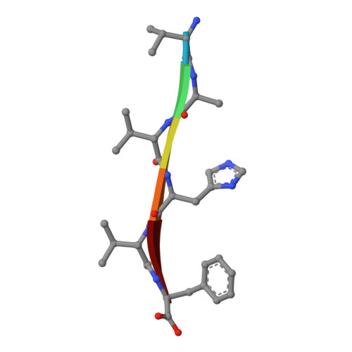
Crystal structures of amyloidogenic segments of human transthyretin.

Saelices, L.Sievers, S.A.Sawaya, M.R.Eisenberg, D.S.

(2018) Protein Sci 27: 1295-1303

  • DOI: https://doi.org/10.1002/pro.3420
  • Primary Citation of Related Structures:  
    6C3F, 6C3G, 6C3S, 6C3T, 6C4O, 6C88

  • PubMed Abstract: 

    Amyloid diseases are characterized by the deposition of proteins in the form of amyloid fibrils, in organs that eventually fail. The development of effective drug candidates follows from the understanding of the molecular processes that lead to protein aggregation. Here, we study amyloidogenic segments of transthyretin (TTR). TTR is a transporter of thyroxine and retinol in the blood and cerebrospinal fluid. When mutated and/or as a result of aging, TTR aggregates into amyloid fibrils that accumulate in organs such as the heart. Recently, we reported two amyloidogenic segments that drive amyloid aggregation. Here, we report the crystal structure of another six amyloidogenic segments of TTR. We found that the segments from the C-terminal region of TTR form in-register steric-zippers with highly-interdigitated, wet interfaces, whereas the β-strand B from the N-terminal region of TTR forms an out-of-register assembly, previously associated with oligomeric formation. Our results contribute fundamental information for understanding the mechanism of aggregation of TTR.


  • Organizational Affiliation

    Departments of Biological Chemistry and Chemistry and Biochemistry, Molecular Biology Institute, Box 951570, UCLA, Howard Hughes Medical Institute, UCLA-DOE Institute, Los Angeles, California, 90095-1570.


Macromolecules

Find similar proteins by:  Sequence   |   3D Structure  

Entity ID: 1
MoleculeChains Sequence LengthOrganismDetailsImage
VAL-ALA-VAL-HIS-VAL-PHE
A, B
6Homo sapiensMutation(s): 0 
Sequence Annotations
Expand
  • Reference Sequence
Small Molecules
Ligands 1 Unique
IDChains Name / Formula / InChI Key2D Diagram3D Interactions
GOL
Query on GOL

Download Ideal Coordinates CCD File 
C [auth B]GLYCEROL
C3 H8 O3
PEDCQBHIVMGVHV-UHFFFAOYSA-N
Experimental Data & Validation

Experimental Data

  • Method: X-RAY DIFFRACTION
  • Resolution: 1.85 Å
  • R-Value Free: 0.178 
  • R-Value Work: 0.171 
  • R-Value Observed: 0.172 
  • Space Group: P 21 21 21
Unit Cell:
Length ( Å )Angle ( ˚ )
a = 11.529α = 90
b = 20.36β = 90
c = 36.729γ = 90
Software Package:
Software NamePurpose
SCALEPACKdata scaling
PHENIXrefinement
PDB_EXTRACTdata extraction
DENZOdata reduction
PHASERphasing

Structure Validation

View Full Validation Report


Entry History & Funding Information

Deposition Data


Funding OrganizationLocationGrant Number
Seventh Framework Programme for ResearchUnited States298559

Revision History  (Full details and data files)

  • Version 1.0: 2018-04-18
    Type: Initial release
  • Version 1.1: 2018-07-18
    Changes: Data collection, Database references
  • Version 1.2: 2018-08-01
    Changes: Data collection, Database references
  • Version 1.3: 2024-03-13
    Changes: Data collection, Database references