6C7Z

Crystal structure of the Q108K:K40L:T51V:R58F mutant of human Cellular Retinol Binding Protein II in complex with synthetic Ligand Julolidine


Experimental Data Snapshot

  • Method: X-RAY DIFFRACTION
  • Resolution: 1.42 Å
  • R-Value Free: 0.256 
  • R-Value Work: 0.222 
  • R-Value Observed: 0.224 

wwPDB Validation   3D Report Full Report


Ligand Structure Quality Assessment 


This is version 1.4 of the entry. See complete history


Literature

A Genetically Encoded Ratiometric pH Probe: Wavelength Regulation-Inspired Design of pH Indicators.

Berbasova, T.Tahmasebi Nick, S.Nosrati, M.Nossoni, Z.Santos, E.M.Vasileiou, C.Geiger, J.H.Borhan, B.

(2018) Chembiochem 19: 1288-1295

  • DOI: https://doi.org/10.1002/cbic.201800050
  • Primary Citation of Related Structures:  
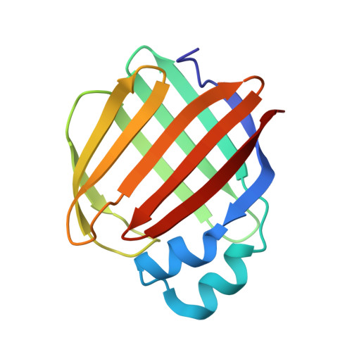
    6C7Z

  • PubMed Abstract: 

    Mutants of human cellular retinol-binding protein II (hCRBPII) were engineered to bind a julolidine retinal analogue for the purpose of developing a ratiometric pH sensor. The design relied on the electrostatic influence of a titratable amino acid side chain, which affects the absorption and, thus, the emission of the protein/fluorophore complex. The ratio of emissions obtained at two excitation wavelengths that correspond to the absorption of the two forms of the protein/fluorophore complex, leads to a concentration-independent measure of pH.


  • Organizational Affiliation

    Department of Chemistry, Michigan State University, East Lansing, MI, 48824, USA.


Macromolecules
Find similar proteins by:  (by identity cutoff)  |  3D Structure
Entity ID: 1
MoleculeChains Sequence LengthOrganismDetailsImage
Retinol-binding protein 2
A, B
133Homo sapiensMutation(s): 4 
Gene Names: RBP2CRBP2
UniProt & NIH Common Fund Data Resources
Find proteins for P50120 (Homo sapiens)
Explore P50120 
Go to UniProtKB:  P50120
PHAROS:  P50120
GTEx:  ENSG00000114113 
Entity Groups  
Sequence Clusters30% Identity50% Identity70% Identity90% Identity95% Identity100% Identity
UniProt GroupP50120
Sequence Annotations
Expand
  • Reference Sequence
Experimental Data & Validation

Experimental Data

  • Method: X-RAY DIFFRACTION
  • Resolution: 1.42 Å
  • R-Value Free: 0.256 
  • R-Value Work: 0.222 
  • R-Value Observed: 0.224 
  • Space Group: P 1
Unit Cell:
Length ( Å )Angle ( ˚ )
a = 29.787α = 90.51
b = 35.947β = 92.45
c = 64.089γ = 113.31
Software Package:
Software NamePurpose
PHENIXrefinement
HKL-3000data reduction
HKL-3000data scaling
PHENIXphasing

Structure Validation

View Full Validation Report


Ligand Structure Quality Assessment 


Entry History & Funding Information

Deposition Data


Funding OrganizationLocationGrant Number
National Institutes of Health/National Institute of General Medical Sciences (NIH/NIGMS)United StatesGM101353

Revision History  (Full details and data files)

  • Version 1.0: 2018-04-25
    Type: Initial release
  • Version 1.1: 2018-07-04
    Changes: Data collection, Database references
  • Version 1.2: 2019-02-20
    Changes: Author supporting evidence, Data collection
  • Version 1.3: 2022-03-23
    Changes: Author supporting evidence, Database references
  • Version 1.4: 2023-10-04
    Changes: Data collection, Refinement description