Insights into the pathogenesis of dominant retinitis pigmentosa associated with a D477G mutation in RPE65.
Choi, E.H., Suh, S., Sander, C.L., Hernandez, C.J.O., Bulman, E.R., Khadka, N., Dong, Z., Shi, W., Palczewski, K., Kiser, P.D.(2018) Hum Mol Genet 27: 2225-2243
- PubMed: 29659842
- DOI: https://doi.org/10.1093/hmg/ddy128
- Primary Citation of Related Structures:
6C7K, 6C7O, 6C7P - PubMed Abstract:
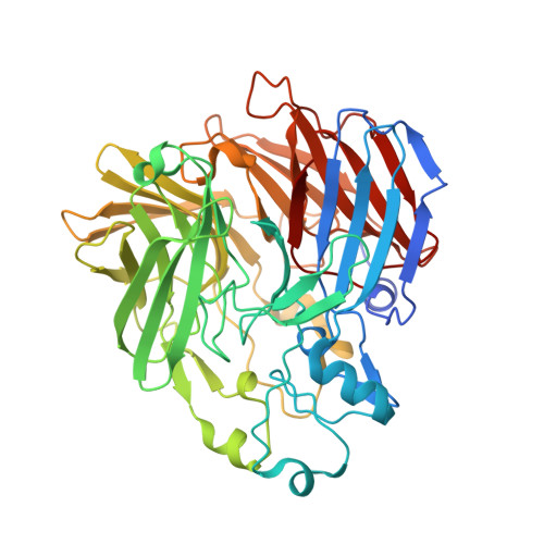
RPE65 is the essential trans-cis isomerase of the classical retinoid (visual) cycle. Mutations in RPE65 give rise to severe retinal dystrophies, most of which are associated with loss of protein function and recessive inheritance. The only known exception is a c.1430G>A (D477G) mutation that gives rise to dominant retinitis pigmentosa with delayed onset and choroidal and macular involvement. Position 477 is distant from functionally critical regions of RPE65. Hence, the mechanism of D477G pathogenicity remains unclear, although protein misfolding and aggregation mechanisms have been suggested. We characterized a D477G knock-in mouse model which exhibited mild age-dependent changes in retinal structure and function. Immunoblot analysis of protein extracts from the eyes of these knock-in mice demonstrated the presence of ubiquitinated RPE65 and reduced RPE65 expression. We observed an accumulation of retinyl esters in the knock-in mice as well as a delay in rhodopsin regeneration kinetics and diminished electroretinography responses, indicative of RPE65 functional impairment induced by the D477G mutation in vivo. However, a cell line expressing D477G RPE65 revealed protein expression levels, cellular localization and retinoid isomerase activity comparable to cells expressing wild-type protein. Structural analysis of an RPE65 chimera suggested that the D477G mutation does not perturb protein folding or tertiary structure. Instead, the mutation generates an aggregation-prone surface that could induce cellular toxicity through abnormal complex formation as suggested by crystal packing analysis. These results indicate that a toxic gain-of-function induced by the D477G RPE65 substitution may play a role in the pathogenesis of this form of dominant retinitis pigmentosa.
Organizational Affiliation:
Department of Pharmacology, School of Medicine, Case Western Reserve University, Cleveland, OH 44106, USA.