6BV2

Crystal structure of porcine aminopeptidase-N with Isoleucine

  • Classification: HYDROLASE
  • Organism(s): Sus scrofa
  • Expression System: Spodoptera frugiperda
  • Mutation(s): No 

  • Deposited: 2017-12-12 Released: 2018-01-17 
  • Deposition Author(s): Chen, L., Lin, Y.-L., Li, F.
  • Funding Organization(s): National Institutes of Health/National Institute Of Allergy and Infectious Diseases (NIH/NIAID)

Experimental Data Snapshot

  • Method: X-RAY DIFFRACTION
  • Resolution: 2.14 Å
  • R-Value Free: 0.257 
  • R-Value Work: 0.213 
  • R-Value Observed: 0.215 

wwPDB Validation   3D Report Full Report


Ligand Structure Quality Assessment 


This is version 2.1 of the entry. See complete history


Literature

The Rational Design of Therapeutic Peptides for Aminopeptidase N using a Substrate-Based Approach.

Joshi, S.Chen, L.Winter, M.B.Lin, Y.L.Yang, Y.Shapovalova, M.Smith, P.M.Liu, C.Li, F.LeBeau, A.M.

(2017) Sci Rep 7: 1424

  • DOI: https://doi.org/10.1038/s41598-017-01542-5
  • Primary Citation of Related Structures:  
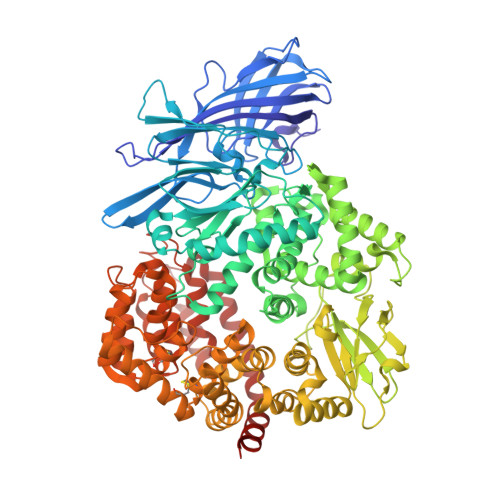
    6BUY, 6BV0, 6BV1, 6BV2, 6BV3, 6BV4

  • PubMed Abstract: 

    The M1 family of metalloproteases represents a large number of exopeptidases that cleave single amino acid residues from the N-terminus of peptide substrates. One member of this family that has been well studied is aminopeptidase N (APN), a multifunctional protease known to cleave biologically active peptides and aide in coronavirus entry. The proteolytic activity of APN promotes cancer angiogenesis and metastasis making it an important target for cancer therapy. To understand the substrate specificity of APN for the development of targeted inhibitors, we used a global substrate profiling method to determine the P1-P4' amino acid preferences. The key structural features of the APN pharmacophore required for substrate recognition were elucidated by x-ray crystallography. By combining these substrate profiling and structural data, we were able to design a selective peptide inhibitor of APN that was an effective therapeutic both in vitro and in vivo against APN-expressing prostate cancer models.


  • Organizational Affiliation

    Department of Pharmacology, University of Minnesota Medical School, Minneapolis, MN, 55455, USA.


Macromolecules
Find similar proteins by:  (by identity cutoff)  |  3D Structure
Entity ID: 1
MoleculeChains Sequence LengthOrganismDetailsImage
Aminopeptidase N902Sus scrofaMutation(s): 0 
Gene Names: ANPEP
EC: 3.4.11.2
UniProt
Find proteins for P15145 (Sus scrofa)
Explore P15145 
Go to UniProtKB:  P15145
Entity Groups  
Sequence Clusters30% Identity50% Identity70% Identity90% Identity95% Identity100% Identity
UniProt GroupP15145
Sequence Annotations
Expand
  • Reference Sequence
Oligosaccharides

Help

Entity ID: 2
MoleculeChains Length2D Diagram Glycosylation3D Interactions
2-acetamido-2-deoxy-beta-D-glucopyranose-(1-4)-2-acetamido-2-deoxy-beta-D-glucopyranose-(1-4)-2-acetamido-2-deoxy-beta-D-glucopyranose
B, C, F
3N-Glycosylation
Glycosylation Resources
GlyTouCan:  G47362BJ
GlyCosmos:  G47362BJ
GlyGen:  G47362BJ
Entity ID: 3
MoleculeChains Length2D Diagram Glycosylation3D Interactions
2-acetamido-2-deoxy-beta-D-glucopyranose-(1-4)-2-acetamido-2-deoxy-beta-D-glucopyranose
D, E, G, H, I
D, E, G, H, I, J
2N-Glycosylation
Glycosylation Resources
GlyTouCan:  G42666HT
GlyCosmos:  G42666HT
GlyGen:  G42666HT
Small Molecules
Ligands 4 Unique
IDChains Name / Formula / InChI Key2D Diagram3D Interactions
NAG
Query on NAG

Download Ideal Coordinates CCD File 
K [auth A],
L [auth A]
2-acetamido-2-deoxy-beta-D-glucopyranose
C8 H15 N O6
OVRNDRQMDRJTHS-FMDGEEDCSA-N
ILE
Query on ILE

Download Ideal Coordinates CCD File 
M [auth A]ISOLEUCINE
C6 H13 N O2
AGPKZVBTJJNPAG-WHFBIAKZSA-N
SO4
Query on SO4

Download Ideal Coordinates CCD File 
N [auth A]
O [auth A]
P [auth A]
Q [auth A]
R [auth A]
N [auth A],
O [auth A],
P [auth A],
Q [auth A],
R [auth A],
S [auth A],
T [auth A],
U [auth A],
V [auth A],
W [auth A]
SULFATE ION
O4 S
QAOWNCQODCNURD-UHFFFAOYSA-L
ZN
Query on ZN

Download Ideal Coordinates CCD File 
X [auth A]ZINC ION
Zn
PTFCDOFLOPIGGS-UHFFFAOYSA-N
Biologically Interesting Molecules (External Reference) 1 Unique
Experimental Data & Validation

Experimental Data

  • Method: X-RAY DIFFRACTION
  • Resolution: 2.14 Å
  • R-Value Free: 0.257 
  • R-Value Work: 0.213 
  • R-Value Observed: 0.215 
  • Space Group: C 1 2 1
Unit Cell:
Length ( Å )Angle ( ˚ )
a = 260.359α = 90
b = 62.923β = 100.51
c = 81.733γ = 90
Software Package:
Software NamePurpose
REFMACrefinement
HKL-2000data reduction
HKL-2000data scaling
PHASERphasing

Structure Validation

View Full Validation Report


Ligand Structure Quality Assessment 


Entry History & Funding Information

Deposition Data


Funding OrganizationLocationGrant Number
National Institutes of Health/National Institute Of Allergy and Infectious Diseases (NIH/NIAID)United StatesR01AI089728

Revision History  (Full details and data files)

  • Version 1.0: 2018-01-17
    Type: Initial release
  • Version 1.1: 2018-02-14
    Changes: Author supporting evidence
  • Version 1.2: 2019-12-18
    Changes: Author supporting evidence
  • Version 2.0: 2020-07-29
    Type: Remediation
    Reason: Carbohydrate remediation
    Changes: Advisory, Atomic model, Data collection, Derived calculations, Refinement description, Structure summary
  • Version 2.1: 2023-10-04
    Changes: Data collection, Database references, Refinement description, Structure summary