6BT6

CTX-M-14 S237A Beta-Lactamase in Complex with a Non-Covalent Tetrazole Inhibitor


Experimental Data Snapshot

  • Method: X-RAY DIFFRACTION
  • Resolution: 1.05 Å
  • R-Value Free: 0.148 
  • R-Value Work: 0.126 
  • R-Value Observed: 0.127 

wwPDB Validation   3D Report Full Report


Ligand Structure Quality Assessment 


This is version 2.1 of the entry. See complete history


Literature

Antibacterial Spectrum of a Tetrazole-Based Reversible Inhibitor of Serine beta-Lactamases.

Pemberton, O.A.Zhang, X.Nichols, D.A.DeFrees, K.Jaishankar, P.Bonnet, R.Adams, J.Shaw, L.N.Renslo, A.R.Chen, Y.

(2018) Antimicrob Agents Chemother 62

  • DOI: https://doi.org/10.1128/AAC.02563-17
  • Primary Citation of Related Structures:  
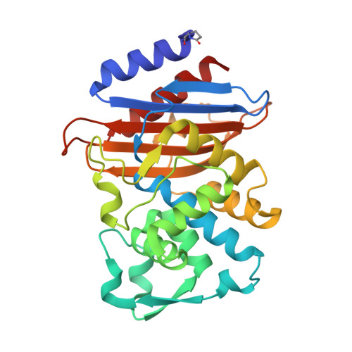
    6BT6, 6BU3

  • PubMed Abstract: 

    CTX-M is the most prevalent family of extended-spectrum β-lactamases. We recently developed a tetrazole-derived noncovalent inhibitor of CTX-M-9. Here, we present the biochemical and microbiological activity of this inhibitor across a representative panel of serine β-lactamases and Gram-negative bacteria. The compound displayed significant activity against all major subgroups of CTX-M, including CTX-M-15, while it exhibited some low-level inhibition of other serine β-lactamases. Complex crystal structures with the CTX-M-14 S237A mutant and CTX-M-27 illustrate the binding contribution of specific active-site residues on the β3 strand. In vitro pharmacokinetic studies revealed drug-like properties and positive prospects for further optimization. These studies suggest that tetrazole-based compounds can provide novel chemotypes for future serine β-lactamase inhibitor discovery.


  • Organizational Affiliation

    Department of Molecular Medicine, University of South Florida, Tampa, Florida, USA.


Macromolecules
Find similar proteins by:  (by identity cutoff)  |  3D Structure
Entity ID: 1
MoleculeChains Sequence LengthOrganismDetailsImage
Beta-lactamase
A, B
263Escherichia coliMutation(s): 1 
Gene Names: blaCTX-M
EC: 3.5.2.6
UniProt
Find proteins for Q9L5C7 (Escherichia coli)
Explore Q9L5C7 
Go to UniProtKB:  Q9L5C7
Entity Groups  
Sequence Clusters30% Identity50% Identity70% Identity90% Identity95% Identity100% Identity
UniProt GroupQ9L5C7
Sequence Annotations
Expand
  • Reference Sequence
Small Molecules
Modified Residues  1 Unique
IDChains TypeFormula2D DiagramParent
PCA
Query on PCA
A, B
L-PEPTIDE LINKINGC5 H7 N O3GLN
Binding Affinity Annotations 
IDSourceBinding Affinity
3GK Binding MOAD:  6BT6 Ki: 1230 (nM) from 1 assay(s)
Experimental Data & Validation

Experimental Data

  • Method: X-RAY DIFFRACTION
  • Resolution: 1.05 Å
  • R-Value Free: 0.148 
  • R-Value Work: 0.126 
  • R-Value Observed: 0.127 
  • Space Group: P 1 21 1
Unit Cell:
Length ( Å )Angle ( ˚ )
a = 45.19α = 90
b = 107.13β = 101.52
c = 47.85γ = 90
Software Package:
Software NamePurpose
iMOSFLMdata reduction
SCALAdata scaling
PHASERphasing
PHENIXrefinement
PDB_EXTRACTdata extraction

Structure Validation

View Full Validation Report


Ligand Structure Quality Assessment 


Entry History & Funding Information

Deposition Data


Funding OrganizationLocationGrant Number
National Institutes of Health/National Institute Of Allergy and Infectious Diseases (NIH/NIAID)United StatesAI103158

Revision History  (Full details and data files)

  • Version 1.0: 2018-06-13
    Type: Initial release
  • Version 1.1: 2019-02-06
    Changes: Data collection, Database references, Structure summary
  • Version 1.2: 2019-02-20
    Changes: Author supporting evidence, Data collection
  • Version 2.0: 2019-12-18
    Changes: Author supporting evidence, Polymer sequence
  • Version 2.1: 2023-10-04
    Changes: Data collection, Database references, Refinement description