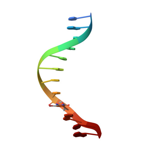
Binding of an antitumor drug to DNA, Netropsin and C-G-C-G-A-A-T-T-BrC-G-C-G.
Kopka, M.L., Yoon, C., Goodsell, D., Pjura, P., Dickerson, R.E.(1985) J Mol Biol 183: 553-563
- PubMed: 2991536
- DOI: https://doi.org/10.1016/0022-2836(85)90171-8
- Primary Citation of Related Structures:
6BNA - PubMed Abstract:
The antitumor antibiotic netropsin has been co-crystallized with a double-helical B-DNA dodecanucleotide of sequence: C-G-C-G-A-A-T-T-BrC-G-C-G, and the structure of the complex has been solved by X-ray diffraction at a resolution of 2.2 A. The structure has been refined independently by Jack-Levitt and Hendrickson-Konnert least-squares methods, leading to a final residual error of 0.257 by the Jack-Levitt approach (0.211 for two-sigma data) or 0.248 by the Hendrickson-Konnert approach, with no significant difference between refined structures. The netropsin molecule displaces the spine of hydration and fits snugly within the minor groove in the A-A-T-T center. It widens the groove slightly and bends the helix axis back by 8 degrees, but neither unwinds nor elongates the double helix. The drug molecule is held in place by amide NH hydrogen bonds that bridge adenine N-3 and thymine O-2 atoms, exactly as with the spine of hydration. The requirement of A X T base-pairs in the binding site arises because the N-2 amino group of guanine would demand impermissibly close contacts with netropsin. It is proposed that substitution of imidazole for pyrrole in netropsin should create a family of "lexitropsins" capable of reading G X C-containing base sequences.