6BDO

Structure of bacterial type II NADH dehydrogenase from Caldalkalibacillus thermarum complexed with a quinone inhibitor HQNO at 2.8A resolution


Experimental Data Snapshot

  • Method: X-RAY DIFFRACTION
  • Resolution: 2.80 Å
  • R-Value Free: 0.268 
  • R-Value Work: 0.227 
  • R-Value Observed: 0.229 

wwPDB Validation   3D Report Full Report


Ligand Structure Quality Assessment 


This is version 1.3 of the entry. See complete history


Literature

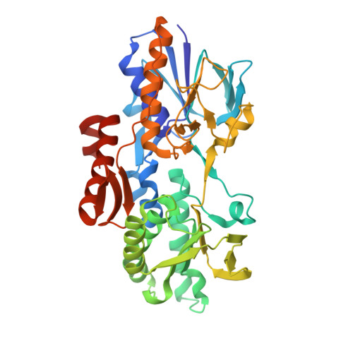
Structure of the NDH-2 - HQNO inhibited complex provides molecular insight into quinone-binding site inhibitors.

Petri, J.Shimaki, Y.Jiao, W.Bridges, H.R.Russell, E.R.Parker, E.J.Aragao, D.Cook, G.M.Nakatani, Y.

(2018) Biochim Biophys Acta 1859: 482-490

  • DOI: https://doi.org/10.1016/j.bbabio.2018.03.014
  • Primary Citation of Related Structures:  
    6BDO

  • PubMed Abstract: 

    Type II NADH:quinone oxidoreductase (NDH-2) is a proposed drug-target of major pathogenic microorganisms such as Mycobacterium tuberculosis and Plasmodium falciparum. Many NDH-2 inhibitors have been identified, but rational drug development is impeded by the lack of information regarding their mode of action and associated inhibitor-bound NDH-2 structure. We have determined the crystal structure of NDH-2 complexed with a quinolone inhibitor 2-heptyl-4-hydroxyquinoline-N-oxide (HQNO). HQNO is nested into the slot-shaped tunnel of the Q-site, in which the quinone-head group is clamped by Q317 and I379 residues, and hydrogen-bonds to FAD. The interaction of HQNO with bacterial NDH-2 is very similar to the native substrate ubiquinone (UQ 1 ) interactions in the yeast Ndi1-UQ 1 complex structure, suggesting a conserved mechanism for quinone binding. Further, the structural analysis provided insight how modifications of quinolone scaffolds improve potency (e.g. quinolinyl pyrimidine derivatives) and suggests unexplored target space for the rational design of new NDH-2 inhibitors.


  • Organizational Affiliation

    Department of Microbiology and Immunology, University of Otago, Dunedin 9054, New Zealand.


Macromolecules
Find similar proteins by:  (by identity cutoff)  |  3D Structure
Entity ID: 1
MoleculeChains Sequence LengthOrganismDetailsImage
FAD-dependent pyridine nucleotide-disulfide oxidoreductaseA [auth B],
B [auth A],
C [auth D],
D [auth C]
405Caldalkalibacillus thermarum TA2.A1Mutation(s): 0 
Gene Names: CathTA2_0279
UniProt
Find proteins for F5L3B8 (Caldalkalibacillus thermarum (strain TA2.A1))
Explore F5L3B8 
Go to UniProtKB:  F5L3B8
Entity Groups  
Sequence Clusters30% Identity50% Identity70% Identity90% Identity95% Identity100% Identity
UniProt GroupF5L3B8
Sequence Annotations
Expand
  • Reference Sequence
Experimental Data & Validation

Experimental Data

  • Method: X-RAY DIFFRACTION
  • Resolution: 2.80 Å
  • R-Value Free: 0.268 
  • R-Value Work: 0.227 
  • R-Value Observed: 0.229 
  • Space Group: P 1 21 1
Unit Cell:
Length ( Å )Angle ( ˚ )
a = 72.847α = 90
b = 114.342β = 91.17
c = 130.061γ = 90
Software Package:
Software NamePurpose
PHENIXrefinement
XDSdata reduction
Aimlessdata scaling
PHASERphasing

Structure Validation

View Full Validation Report


Ligand Structure Quality Assessment 


Entry History & Funding Information

Deposition Data


Funding OrganizationLocationGrant Number
Health Research Council (HRC)New Zealand--

Revision History  (Full details and data files)

  • Version 1.0: 2018-05-16
    Type: Initial release
  • Version 1.1: 2020-01-01
    Changes: Author supporting evidence
  • Version 1.2: 2023-03-29
    Changes: Database references, Structure summary
  • Version 1.3: 2023-10-25
    Changes: Data collection, Refinement description