6ALQ

VioC L-arginine hydroxylase bound to Fe(II), L-arginine, and succinate


Experimental Data Snapshot

  • Method: X-RAY DIFFRACTION
  • Resolution: 1.67 Å
  • R-Value Free: 0.200 
  • R-Value Work: 0.181 
  • R-Value Observed: 0.182 

wwPDB Validation   3D Report Full Report


Ligand Structure Quality Assessment 


This is version 1.4 of the entry. See complete history


Literature

Visualizing the Reaction Cycle in an Iron(II)- and 2-(Oxo)-glutarate-Dependent Hydroxylase.

Mitchell, A.J.Dunham, N.P.Martinie, R.J.Bergman, J.A.Pollock, C.J.Hu, K.Allen, B.D.Chang, W.C.Silakov, A.Bollinger, J.M.Krebs, C.Boal, A.K.

(2017) J Am Chem Soc 139: 13830-13836

  • DOI: https://doi.org/10.1021/jacs.7b07374
  • Primary Citation of Related Structures:  
    6ALM, 6ALN, 6ALO, 6ALP, 6ALQ, 6ALR

  • PubMed Abstract: 

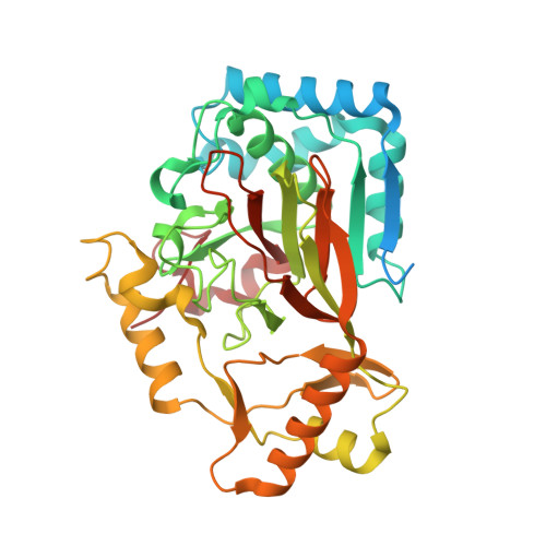
    Iron(II)- and 2-(oxo)-glutarate-dependent oxygenases catalyze diverse oxidative transformations that are often initiated by abstraction of hydrogen from carbon by iron(IV)-oxo (ferryl) complexes. Control of the relative orientation of the substrate C-H and ferryl Fe-O bonds, primarily by direction of the oxo group into one of two cis-related coordination sites (termed inline and offline), may be generally important for control of the reaction outcome. Neither the ferryl complexes nor their fleeting precursors have been crystallographically characterized, hindering direct experimental validation of the offline hypothesis and elucidation of the means by which the protein might dictate an alternative oxo position. Comparison of high-resolution X-ray crystal structures of the substrate complex, an Fe(II)-peroxysuccinate ferryl precursor, and a vanadium(IV)-oxo mimic of the ferryl intermediate in the l-arginine 3-hydroxylase, VioC, reveals coordinated motions of active site residues that appear to control the intermediate geometries to determine reaction outcome.


  • Organizational Affiliation

    Department of Biochemistry and Molecular Biology, The Pennsylvania State University , University Park, Pennsylvania 16802, United States.


Macromolecules
Find similar proteins by:  (by identity cutoff)  |  3D Structure
Entity ID: 1
MoleculeChains Sequence LengthOrganismDetailsImage
Alpha-ketoglutarate-dependent L-arginine hydroxylase394Streptomyces vinaceusMutation(s): 0 
Gene Names: vioC
EC: 1.14.11.41
UniProt
Find proteins for Q6WZB0 (Streptomyces vinaceus)
Explore Q6WZB0 
Go to UniProtKB:  Q6WZB0
Entity Groups  
Sequence Clusters30% Identity50% Identity70% Identity90% Identity95% Identity100% Identity
UniProt GroupQ6WZB0
Sequence Annotations
Expand
  • Reference Sequence
Experimental Data & Validation

Experimental Data

  • Method: X-RAY DIFFRACTION
  • Resolution: 1.67 Å
  • R-Value Free: 0.200 
  • R-Value Work: 0.181 
  • R-Value Observed: 0.182 
  • Space Group: C 1 2 1
Unit Cell:
Length ( Å )Angle ( ˚ )
a = 80.844α = 90
b = 66.751β = 109.27
c = 62.922γ = 90
Software Package:
Software NamePurpose
XSCALEdata scaling
REFMACrefinement
PDB_EXTRACTdata extraction
PHASERphasing

Structure Validation

View Full Validation Report


Ligand Structure Quality Assessment 


Entry History & Funding Information

Deposition Data


Funding OrganizationLocationGrant Number
National Institutes of Health/National Institute of General Medical Sciences (NIH/NIGMS)United StatesGM119707

Revision History  (Full details and data files)

  • Version 1.0: 2017-09-06
    Type: Initial release
  • Version 1.1: 2017-09-27
    Changes: Author supporting evidence
  • Version 1.2: 2017-10-11
    Changes: Database references
  • Version 1.3: 2020-01-01
    Changes: Author supporting evidence
  • Version 1.4: 2024-03-13
    Changes: Data collection, Database references